Institution: University of Texas MD Anderson Cancer Center
Additional authors:Tariq Muzzafar, MD
Session: Erythroleukemia and megakaryoblastic AML and mimics
HISTORY
Patient is a 50 year old male with no significant past medical history presented with two week history of fatigue, weakness, nose bleeding, weight loss, fevers and sweats. He went to the emergency room and was admitted directly to the intensive care unit in view or his clinical state. He was treated with idarubicin and ara-C 3+7 but had persistent disease. He was subsequently given high-dose ara-C and Mylotarg (x1) with refractory disease and liver toxicity. He presented to UTMDACC for treatment options 5 months later. CBC: WBC 4.0, Hgb 9.1, MCV 90, Platelet 3, Neutrophils 28%, Lymphs 63%, Monos 2%, Eos 1%, Bands 1%, Other unclassified cells = 5%
DETAILS
Bone marrow core biopsy (Formalin fixed after decalcification):
90% cellularity, sheets of immature cells replacing 90% of total cellularity; normal hematopoietic cells decreased profoundly
Bone marrow aspirate smears:
93% blasts; blasts are predominantly large, with moderately dispersed chromatin, multiple distinct to prominent nucleoli, abundant, basophilic, agranular cytoplasm and pronounced pseudopod formation
Myeloperoxidase: Blasts are negative
Butyrate esterase: Blasts are negative
IMMUNOHISTOCHEMISTRY AND FLOW CYTOMETRY
Blasts are positive for CD13, CD34, CD36, CD41, CD61
CYTOGENETIC FINDINGS
Karyotype: 46,XY,del(9)(q22)[17] ;46,XY,del(9)(q22)[cp2] 46,XY[1]
MOLECULAR FINDINGS
NEGATIVE for PML-RARA short or long form fusion transcript, AML1-ETO fusion transcript, CBFb-MYH11 fusion transcript, mutations in FLT3 (internal tandem duplication or codon 835/836), K-ras and N-ras oncogenes (codons 12, 13 and 61)
INTERESTING FEATURES
Acute megakaryoblastic leukemia is a rare disease entity. We performed a study that demonstrated that only a small minority (10%) of cases with morphologic / immunophenotypic features of megakaryocytic differentiation can be classified as megakaryoblastic leukemia using WHO classification. Most cases of megakaryoblastic leukemia currently fit diagnostic criteria for acute myeloid leukemia with myelodysplasia-related changes. In our series, 12 (24%) cases evolved from a pre-existing myeloproliferative neoplasm, a finding not commonly emphasized. Megakaryoblastic differentiation occurs in a spectrum of hematologic neoplasms, with complex karyotype being a common feature. However; survival is poor in all instances. In this case, there was no antecedent history and presentation was acute, thus ruling out progression from underlying myeloid neoplasm.
PROPOSED DIAGNOSIS
Acute megakaryoblastic leukemia
CONSENSUS DIAGNOSIS
Acute megakaryoblastic leukemia
| Bone marrow core biopsy 1 |  |
| Bone marrow core biopsy 2 | 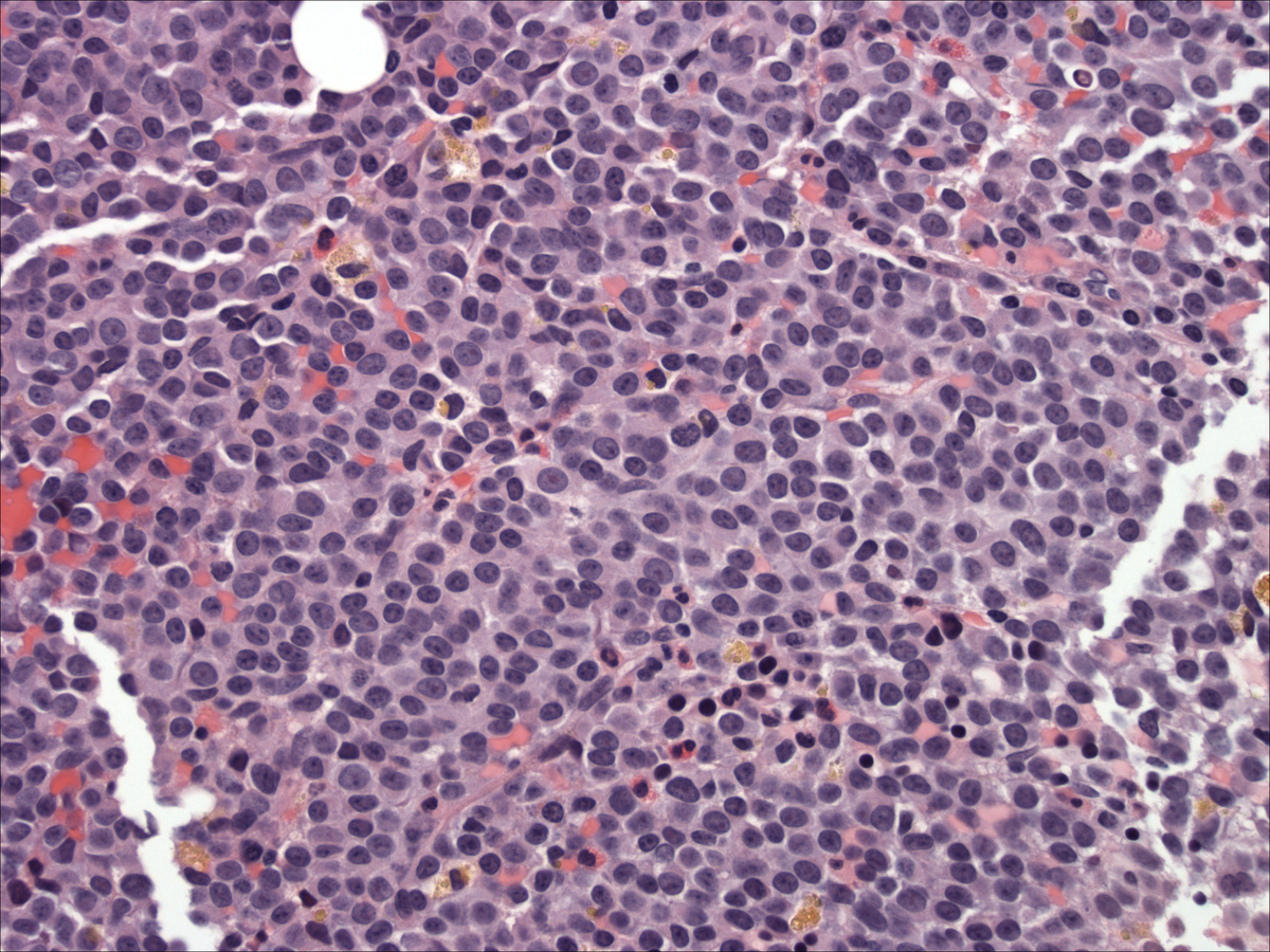 |
| Bone marrow aspirate | 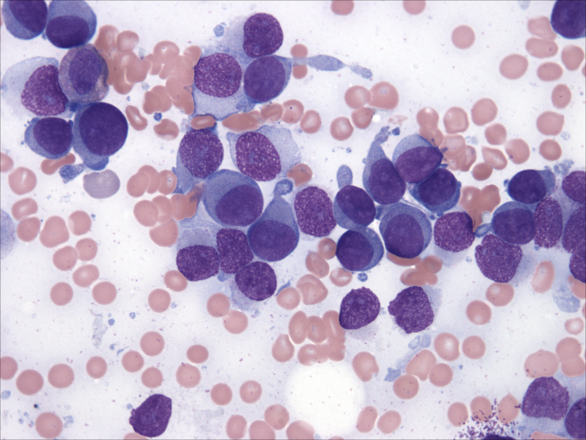 |
| Bone marrow aspirate: myeloperoxidase | 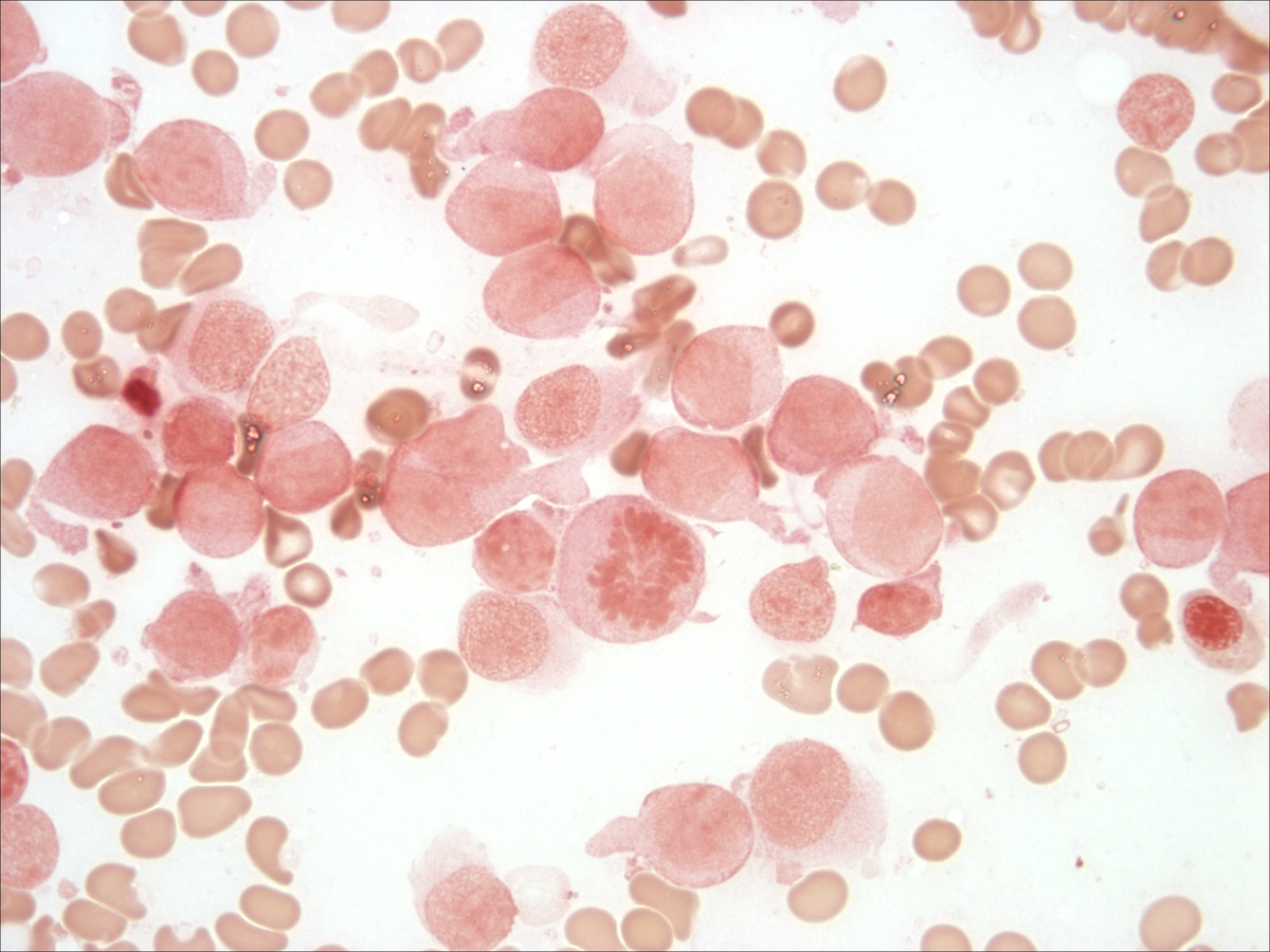 |
| Bone marrow aspirate: butyrate | 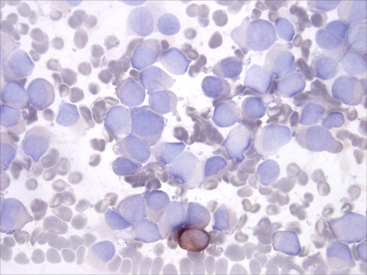 |