Institution: University of Florida
Additional authors:Robert W. Allan, MD, Christopher M. Carter, MD, Mathew R. Simmons, MD, Ying Li, MD, PhD
Session: Extramedullary manifestations of myeloid neoplasms
HISTORY
A 32 year old female with pancytopenia and right maxillary mass. CBC: RBC 2.93, HGB 8.6, HCT 25.2, WBC 3.1, PLT 19, MCV 86.2. Peripheral blood sample was submitted for flow cytometric analysis, followed by biopsy of right maxillary mass and bone marrow biopsy.
DETAILS
The PB smears showed anemia with mild anisopoikilocytosis and mild leukopenia. Increased numbers of immature monocytic cells were seen (~60% of leukocytes). They had immature nuclear chromatin, prominent nucleoli, and a moderate amount of basophilic cytoplasm with occasional small pink granules. The monoblasts were variably NSE-positive and MPO-negative by cytochemical stains.
Histologic sections of maxillary mass biopsies showed a dense atypical mononuclear cell infiltrate composed of intermediate sized cells with fine nuclear chromatin, irregular nuclear contours and prominent nucleoli. Immunohistochemistry showed that the neoplastic cells are lysozyme+, CD4+, CD68+, CD43+, CD123-, myeloperoxidase- and CD56-. The bone marrow touch preparations contained scant cellularity, but the cells present consisted of large blasts with prominent nucleoli and blue cytoplasm. The decalcified and formalin fixed bone marrow core biopsy showed complete replacement by immature-appearing cells with open nuclear chromatin, prominent nucleoli and ample cytoplasm.IMMUNOHISTOCHEMISTRY AND FLOW CYTOMETRY
FLOW CYTOMETRIC ANALYSIS:
a. PERIPHERAL BLOOD: The myeloid blasts were positive for CD4, CD56, CD11c, HLA-DR, CD123, CD13 and CD33, with dim CD15 and dim CD13, without CD34, CD117 or CD11b b. MAXILLARY MASS: Targeted FCM analysis showed the presence of a population of myeloid blasts similar to that seen in the bloodIMMUNOHISTOCHEMISTRY:a. Immunohistochemistry performed on the maxillary biopsy show that the neoplastic cells are lysozyme+, CD11c+, CD4+, CD68+, CD43+, CD123-, myeloperoxidase-, TCL1-, and CD56-. b. Immunohistochemistry performed on the bone marrow core biopsy show that the neoplastic cells are lysozyme+, CD4+ (variable), CD68+, myeloperoxidase- and CD56-. Scattered cells with weak CD123 immunoreactivity were noted.CYTOGENETIC FINDINGS
a. Peripheral Blood Karyotype: 46,XX,t(9;11)(p22;q23)[12]/46,XX[8]
b. Bone Marrow: Interphase FISH study was POSITIVE for MLL gene locus rearrangement. An abnormal population of cells bearing an MLL gene locus rearrangement was observed in ~94.3% of the cells examined from the sample, consistent with the previously identified 9/11 translocation in the chromosome study conducted on PB.INTERESTING FEATURES
1. Concurrent extramedullary myeloid tumor (myeloid sarcoma).
2. The coexpression of CD4, CD56 and CD123, without CD34 or CD117 by flow cytometry suggested plasmacytoid dendritic cell differentiation.3. Differences in expression of CD123 and CD56 between flow cytometry and IHC.PROPOSED DIAGNOSIS
ACUTE MYELOID LEUKEMIA WITH MONOCYTIC DIFFERENTIATION, WITH MLL GENE REARRANGEMENT, AND CONCURRENT EXTRAMEDULLARY MYELOID TUMOR (MYELOID SARCOMA).
CONSENSUS DIAGNOSIS
Acute myeloid leukemia with t(9;11)(p22;q22); MLLT3-MLL, involving bone marrow and right maxillary mass (myeloid sarcoma)
| Peripheral blood | 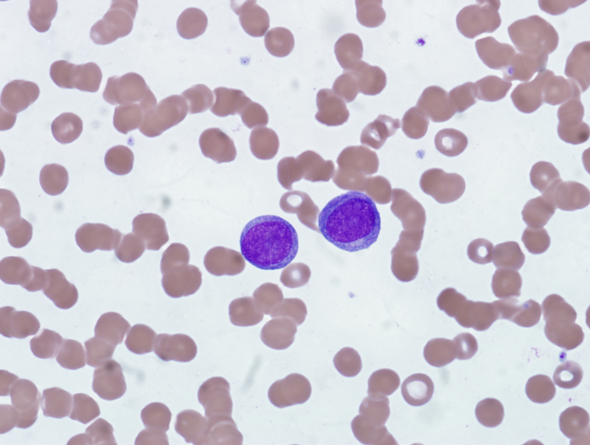 |
| Cytospin preparation of peripheral blood sample | 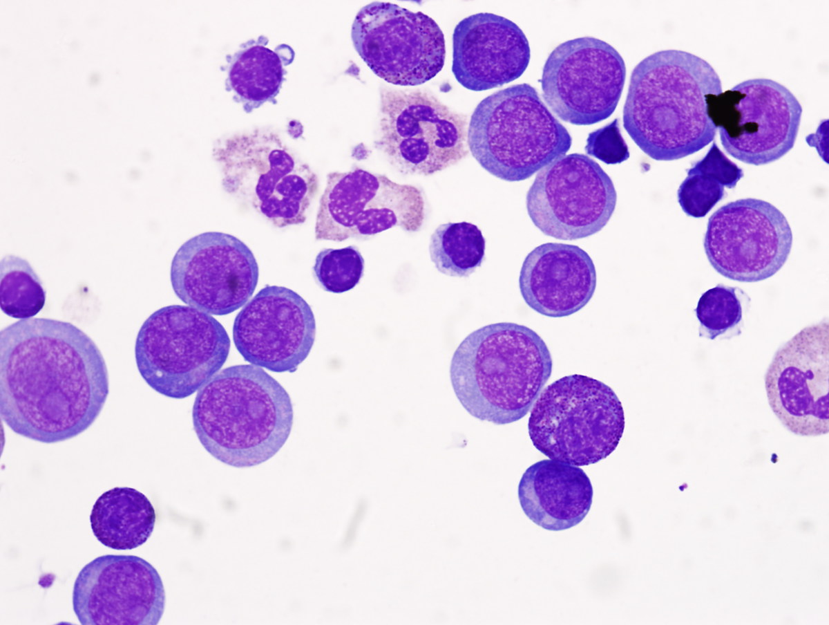 |
| NSE peripheral blood sample |  |
| Maxillary mass biopsy (low power) |  |
| Maxillary mass biopsy (high power) |  |
| Bone marrow biopsy (low power) |  |
| Bone marrow biopsy (high power) |  |
| IHC lysozyme - bone marrow |  |
| IHC Myeloperoxidase - bone marrow |  |
| IHC lysozyme - maxillary biopsy | 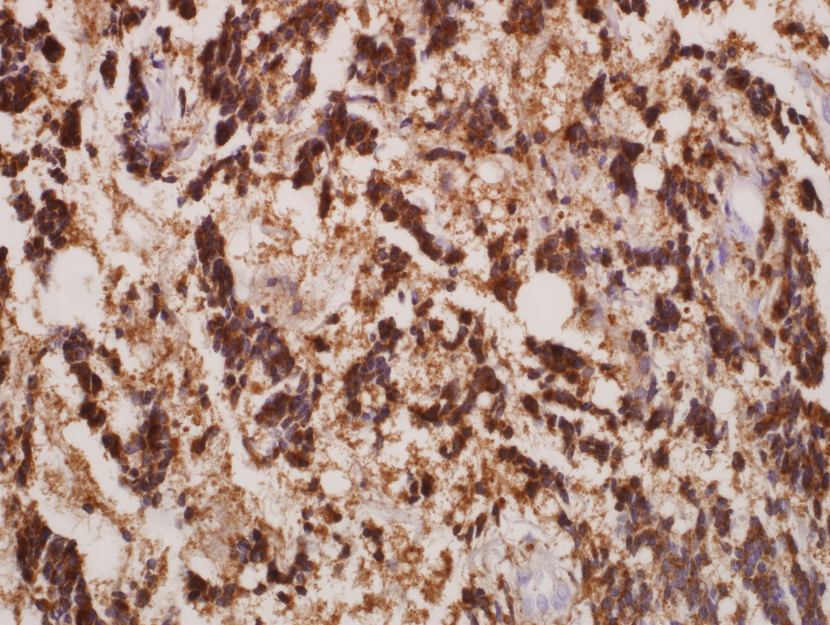 |
| IHC CD43 - maxillary biopsy |  |
| Flow cytometric analysis of PB sample showing the abnormal blasts (red) |  |
| Flow cytometric analysis of PB sample showing the phenotype of abnormal blasts (red) |  |