Institution: UT MD Anderson Cancer Center
Session: AML with recurrent genetic mutations Part II
HISTORY
Patient was a 26-year-old female who in 03/2012 developed chronic bronchitis refractory to antibiotics, low-grade fevers, fatigue, dizziness, and headaches, ecchymotic areas on skin. WBC 28.7, Hgb 9.9, MCV 108.5, Platelet 81, Neutrophils 40%, Lymphs 28%, Monos 11%, Basos 1%, Bands 9%, Metas 6%, Myelo 2%, Blasts 3%
Bone marrow work-up (04/2010 and 05/2012) interpreted as: chronic myelomonocytic leukemia (CMML-2); 11% blasts
Clinician recommended lenilidomide + azacitidine. To enroll on protocol, she needed to be on two forms of contraception. She returned after 4 weeks; over this time she received PRBC (x 5 units) over 7 days but no platelets. Her gums became swollen, and tender and bled easily. WBC: 39.7, HGB: 9.4, MCV: 90, Platelet: 54, with 41% blasts
Bone marrow work-up (06/2012) interpreted as acute myeloid leukemia (AML) evolving from chronic myelomonocytic leukemia.
She received induction chemotherapy with clofarabine, idarubicin, and cytarabine with complete remission and one cycle of consolidation. She had a matched unrelated donor stem cell transplant on 09/2012, post transplant course was complicated by pneumonia, CMV antigenemia, severe mucositis, hematuria with positive BK virus in the urine. She died in 1/2013 due to adult respiratory distress syndrome and respiratory failure.
DETAILS
05/2012 Peripheral blood film: Marked leukocytosis composed predominantly of left-shifted granulocytes with dysplastic features (abnormal nuclear lobation and hypogranular cytoplasm), absolute monocytosis, 5% blasts; severe macrocytic, normochromic anemia with RBCs showing mild to moderate anisopoikilocytosis; moderate thrombocytopenia with occasional hypogranular forms Core biopsy: 100% cellularity; M:E ratio increased, megakaryocytes dysplastic Aspirate smears: trilineage dysplasia, 21% monocytes, cytoplasmic vacuolization prominent, 11% (blasts + promonocytes); butyrate esterase: positive in 30% of cells, rare ring sideroblasts 06/2012 Clot: 100% cellularity, megakaryocytes have atypical features Aspirate smears: 82% blasts; blasts are large with dispersed chromatin, prominent nuclear membrane folds, occasional prominent nucleoli, moderate to abundant cytoplasm with fine granularity; no Auer rods identified; occasional immature monocytes and dysplastic neutrophils also present
IMMUNOHISTOCHEMISTRY AND FLOW CYTOMETRY
Flow cytometry (05/2012) Monocytosis (12 %) with an altered immunophenotype(CD45dec, CD13+, CD33+, CD64+, CD14+, HLADRdec, CD123dec, CD15inc) CD34+ myeloblasts are 0.02% with an altered immunophenotype CD38dec) Abnormal patterns or abnormal antigen expression in myeloid cells Hematogones absent Flow cytometry(06/2012): 67% aberrant cells in CD45(dim) gate POSITIVE: CD33, CD38 (partial), CD49d, CD64 (subset), CD117 (subset), CD123, CD184 (partial), TdT (partial), HLA-DR (partial), and myeloperoxidase NEGATIVE: CD34, CD14, CD2, cytoCD3, CD5, CD7, CD10, CD19, CD41, and CD56
CYTOGENETIC FINDINGS
05/2012
Karyotype:
47,XX,+21[11]
46,XX[9]
06/11/2012
48,XX,+2,6,XX[19]
4% cells have trisomy 21 (FISH)
MOLECULAR FINDINGS
POSITIVE: mutation in NPM1 (exon 12), NRAS (codon 12), DNMT3A (exon 23 codon 882) NEGATIVE: mutations in KRAS, FLT3 (internal tandem duplication or codon 835/836), KIT (exon 17), mutation or alteration detected in CEBPA, IDH1 (exon 4, codons 87 to 138), IDH2 (exon 4), codon 617 of JAK2
INTERESTING FEATURES
Acute myeloid leukemia with mutated NPM1 is strongly correlated with monocytic features; 80-90% acute monocytic leukemias are reported to have NPM1 mutations. However, when these present in evolving phase; bone marrow blast percentage can be less than 20% and thus the case meets criteria for CMML but not acute myeloid leukemia. Patients are stratified as per IPSS scoring and treatment may be delayed. In the meantime, the leukemic clone proliferates and blast percentage rises rapidly and in a few weeks, patient presents as a florid AML with monocytic features. If a patient presents with 11% blast count acutely with no prior antecedent hematologic disorder, it is highly likely s(he) has a high risk myeloid neoplasm which could very well be an AML in evolution (impending AML). This possibility should be considered especially if the patient is young, since CMML is a disease of elderly people with median age at diagnosis being 65-75 years. Blasts in this case were negative for CD34 as is characteristic for AML with mutated NPM. Additional mutations present in this case were NRAS (codon 12), DNMT3A (exon 23 codon 882) Leukemic cells demonstrated rapid clonal selection over a 4 week period; cells with +21 decreased from 55% to 5% and cells with diploid karyotype decreased from 45% to 95% as assessed by karyotypic analysis. The diploid cells presumably had the characteristic mutations that enabled them to proliferate at the expense of the cells with +21. Myeloid neoplasms may originate from heterogeneous clonal populations with more than one clonal population present at inception. The clonal populations with survival advantage would displace other clones and hence the disease would evolve over time with a changing clinical profile and eventual resistance to chemotherapeutic drugs. This phenomenon would need to considered in developing targeted chemotherapeutic drugs, since the target may be a moving target.
PROPOSED DIAGNOSIS
Acute myeloid leukemia with mutated NPM1 presenting in evolving (impending) phase with 11% blasts
CONSENSUS DIAGNOSIS
Acute myeloid leukemia with mutations in NPM1 (exon 12), NRAS (codon 12), DNMT3A (exon 23, codon 882), and trisomy 21
| Bone marrow core biopsy 1 (5/2012) | 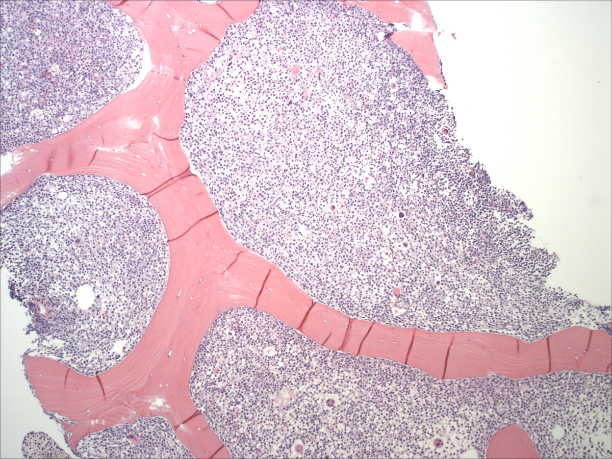 |
| Bone marrow aspirate (5/2012) | 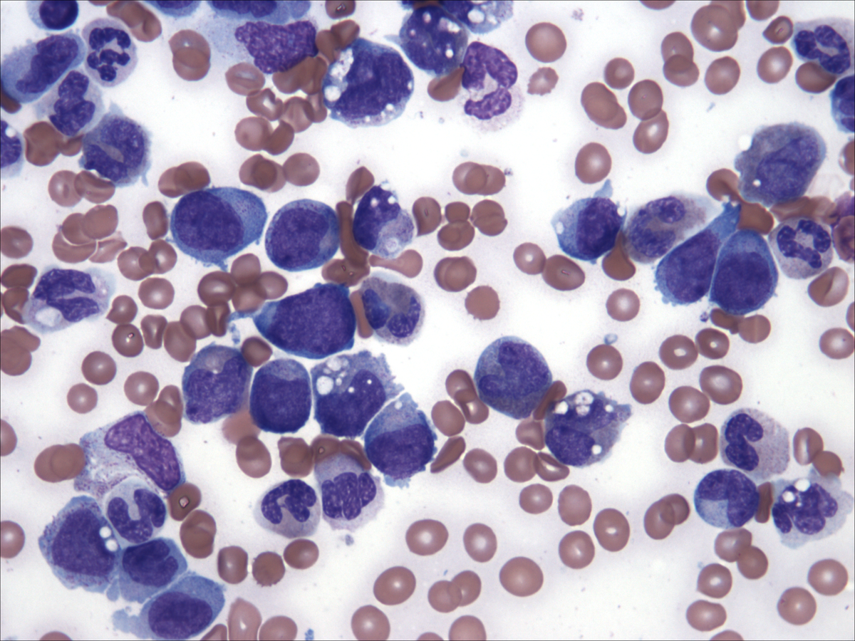 |
| Bone marrow aspirate: butyrate (05/2012) | 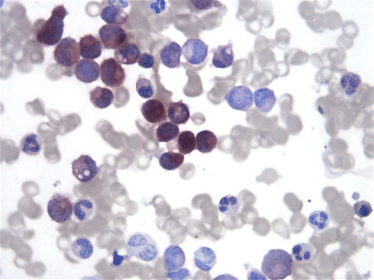 |
| Peripheral blood (05/2012) | 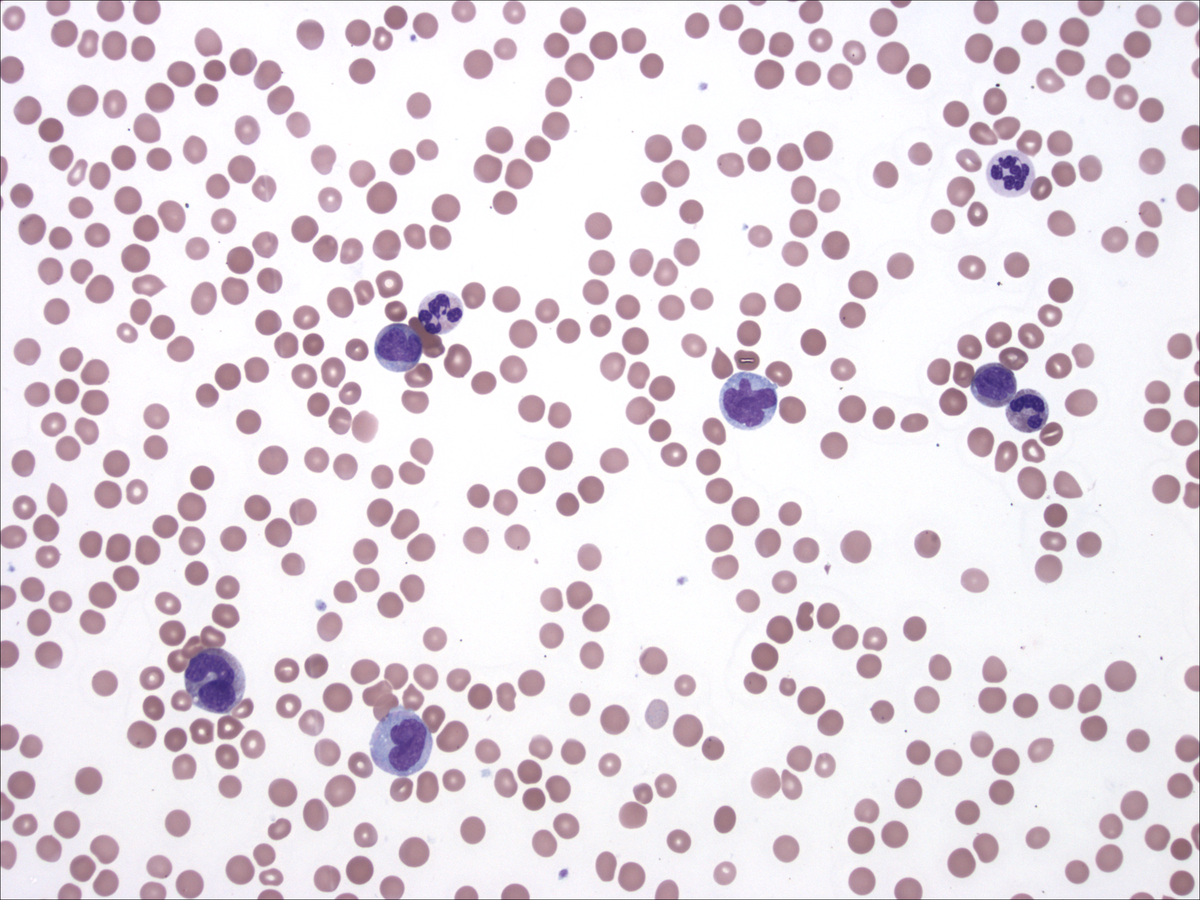 |
| Bone marrow clot (06/2012) | 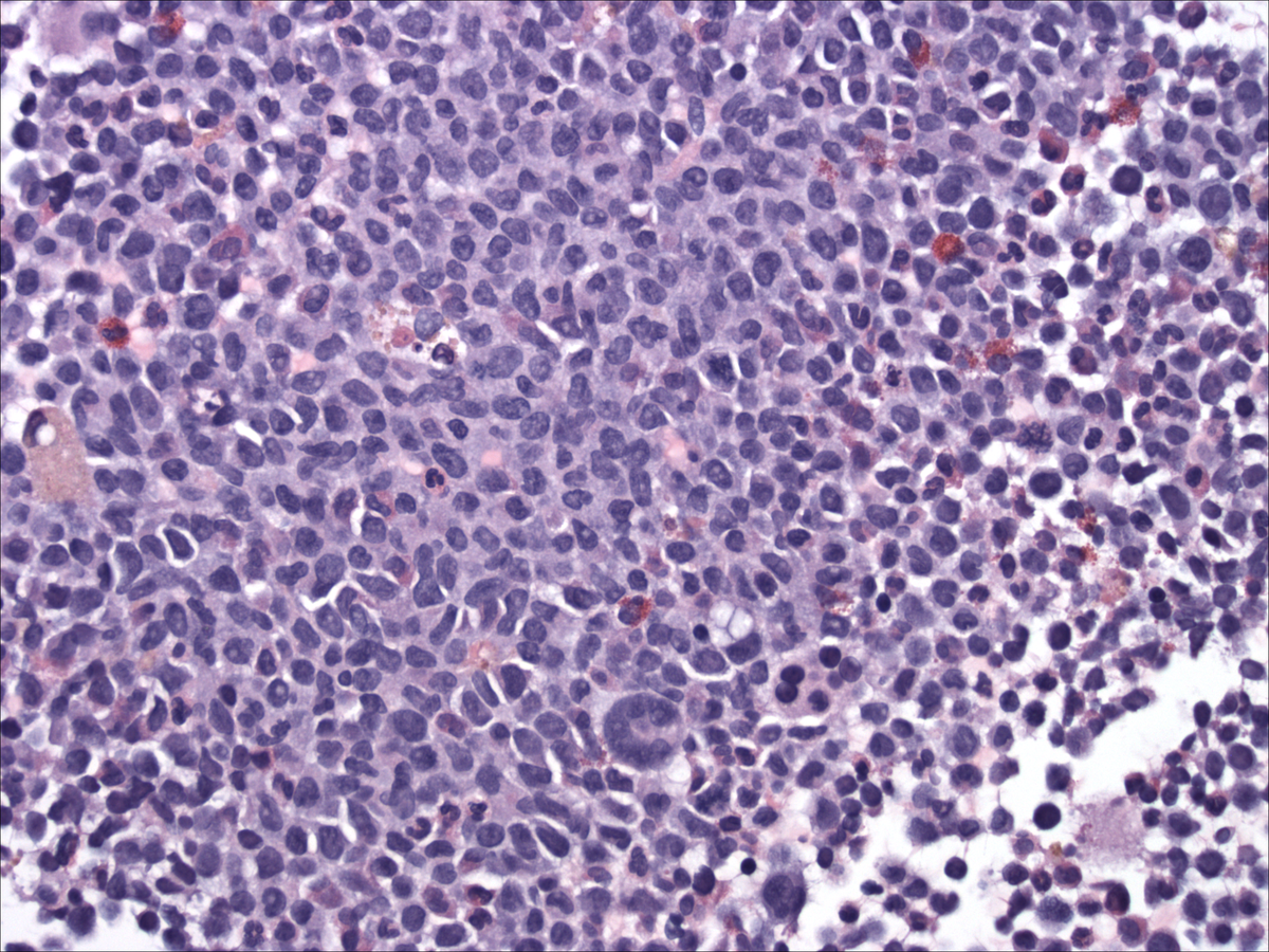 |
| Bone marrow aspirate (06/2012) | 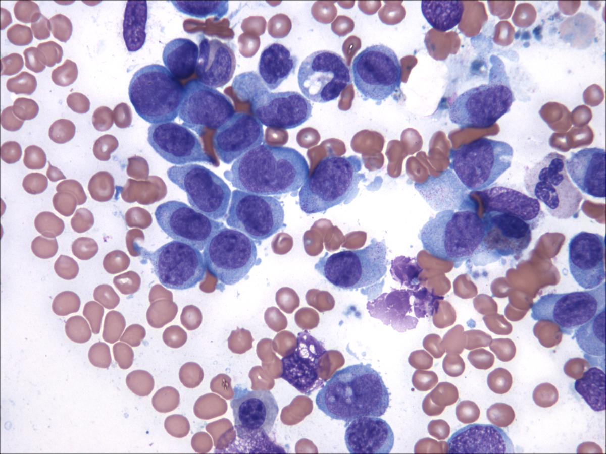 |