Institution: University of Florida
Additional authors:Mathew Simmons, MD, Ying Li, MD, PhD
Session: B Lymphoblastic Leukemia/Lymphoma
HISTORY
A 64-year-old male with history of AML-M5b, diagnosed in 2008 with normal cytogenetics, currently enrolled in Oxi trial. The leukemia relapsed in 2011 with persistent disease through 2012. Multiple bone marrow biopsies showed increased CD34+/CD117+ myeloid blasts (that variably ranged from 4-9%) and was thought to represent underlying myelodysplastic syndrome. A bone marrow biopsy in September 2012 showed increased myeloid blasts (7-9%) and concurrent cytogenetic analysis showed an abnormal karyotype with chromosome 11 long arm interstitial deletion in two of twenty metaphase cells examined. A bone marrow biopsy in January 2012 showed increased myeloid blasts (5%). Concurrent cytogenetic analysis showed three abnormal cell populations identified among nine of twenty metaphase cells examined by conventional cytogenetic techniques. Two of the three abnormal cell populations, represented by six of the nine abnormal metaphase cells, were characterized to include trisomy 8 and a structurally altered chromosome 9, either as an isochromosome 9 long arm or as a ring chromosome 9. The third and apparently unrelated abnormal cell population, represented by three of the nine abnormal metaphase cells, was characterized to include a derivative chromosome 18 that includes a chromosome 1 long arm segment translocated to its long arm. The current bone marrow sample was performed in February 2013
DETAILS
The bone marrow aspirate smears were aspicular, markedly hemodiluted, and suboptimal for evaluation. The touch preparations were similarly inadequate. There were scattered blasts with large nuclei, fine chromatin, and prominent nucleoli with agranular cytoplasm. Occasional nucleated blood cells were seen. The cytospin preparation showed increased blasts, with large nuclei, prominent nucleoli and ample basophilic cytoplasm. The decalcified and formalin fixed bone marrow core biopsy was small and fragmented and composed predominantly of cortical bone and periosteum. There was a very small area of crushed blue cells with increased hemosiderin deposition but no active hematopoietic elements were identified.
IMMUNOHISTOCHEMISTRY AND FLOW CYTOMETRY
An abnormal population of B-lymphoid blasts was identified with expression of CD20, CD19, CD10, CD22, HLA-DR, CD38, CD58 and TdT with variable expression of CD34 and no expression of CD45. The B-lymphoid blasts comprised 10.5% of the cells in the sample.
CYTOGENETIC FINDINGS
KARYOTYPE: 47,XY,+8,i(9)(q10)[cp5]/48,XY,+8,r(9)(p24q34),+21[cp2]/46,XY[13]
INTERPHASE FISH: POSITIVE for trisomy 8 (D8Z1)INTERESTING FEATURES
Evolution of new cytogenetic abnormalities and abnormal B-lymphoid blasts in a patient with history of acute myeloid leukemia.
Although the cytsopin is not reliable for assessment of blast morphology, the blasts show monocytic features despite the immature B-cell phenotype detected by flow cytometryPROPOSED DIAGNOSIS
Abnormal population of B-lymphoid blasts detected. This may represent clonal evolution of underlying stem cell disease (high grade MDS/evolving acute lymphoblastic leukemia)
No evidence of acute myeloid leukemia.CONSENSUS DIAGNOSIS
Therapy related abnormal population of B-lymphoid blasts
No evidence of acute myeloid leukemia.| Bone marrow aspirate | 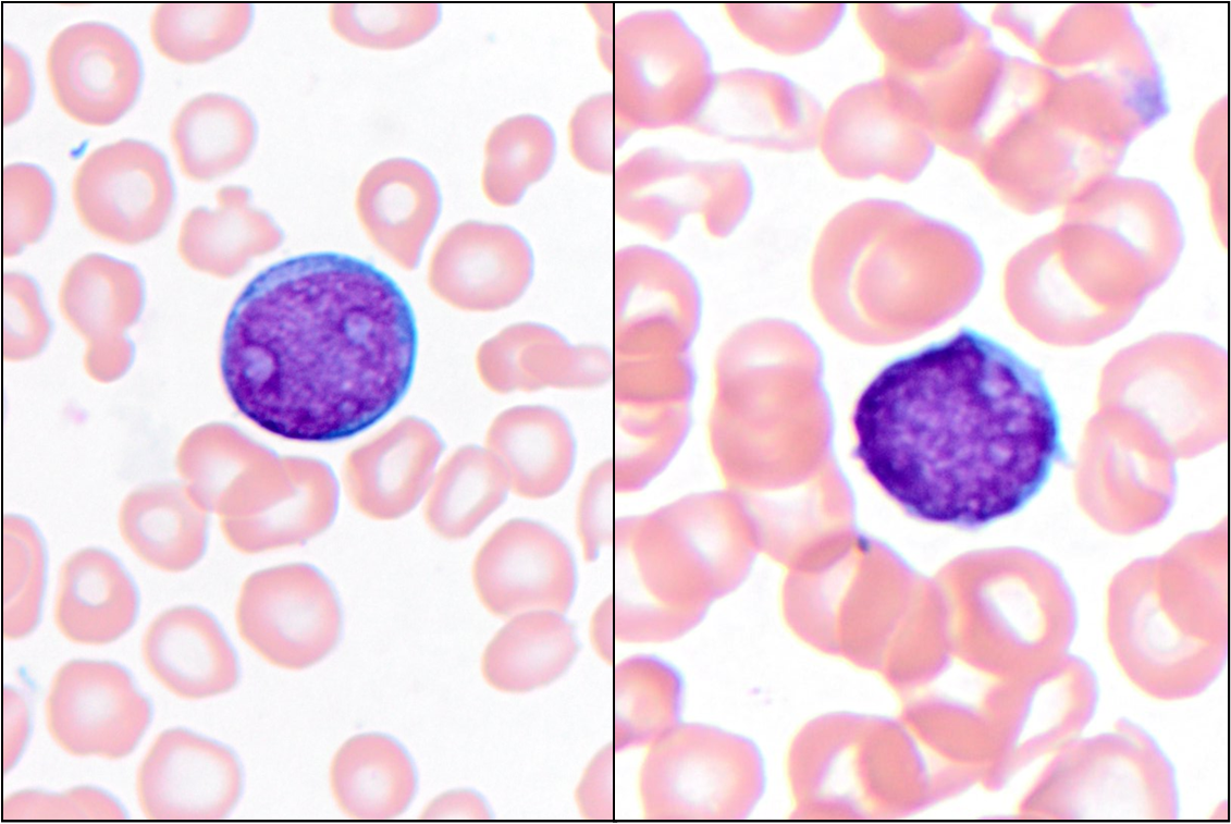 |
| Bone marrow cytospin | 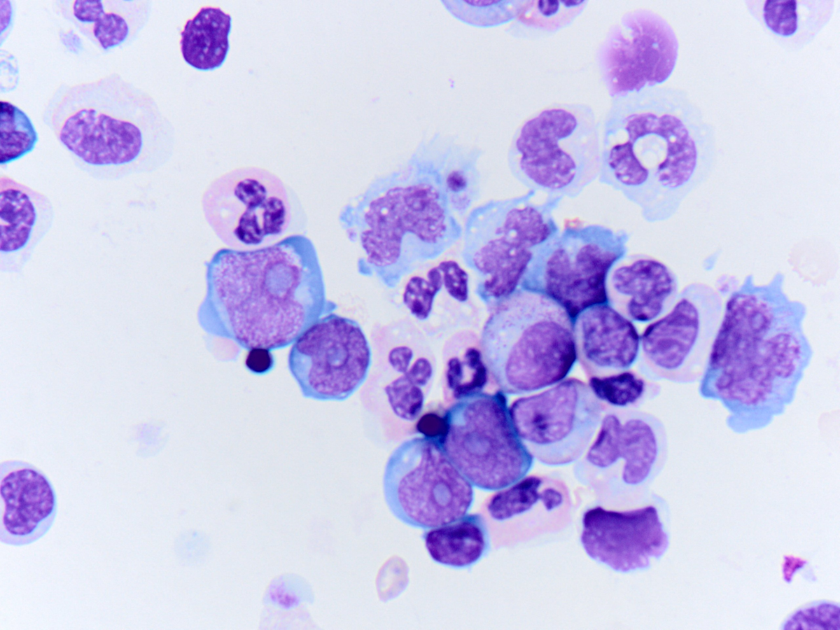 |
| Flow cytometric analysis of BM aspirate with the B-lymphoid blasts (aqua). Myeloid blasts are shown in (yellow) | 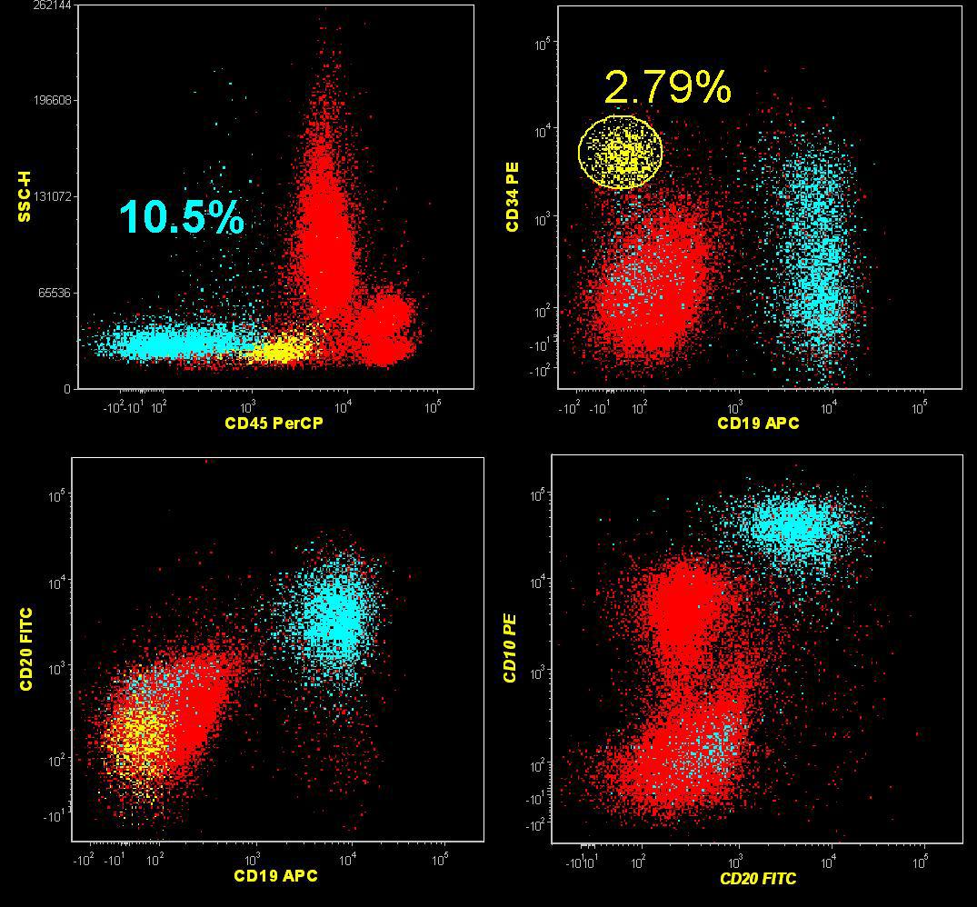 |
| Flow cytometric analysis of BM aspirate with the B-lymphoid blasts (aqua). Myeloid blasts are shown in (yellow) | 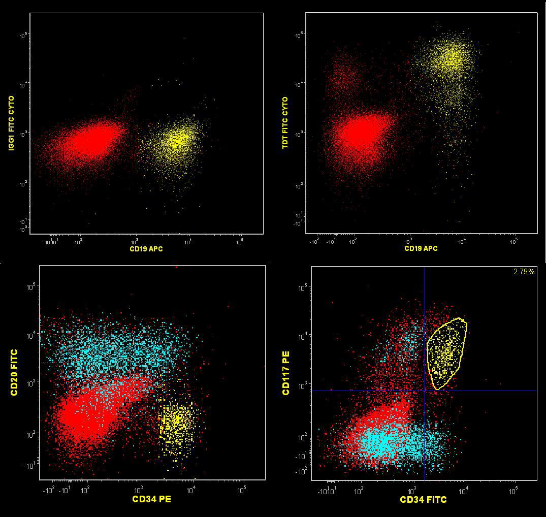 |