Institution: MGH
Additional authors:Robert P. Hasserjian
Session: Erythroleukemia and megakaryoblastic AML and mimics
HISTORY
A 75 year old man with a long-standing history of chronic lymphocytic leukemia treated with chemotherapy (including cladribine, cyclophosphamide, vincristine, and fludarabine) presented with progressive thrombocytopenia. A bone marrow biopsy revealed a therapy-related myelodysplastic syndrome with 15% blasts. He was treated with 5-azacitidine with no improvement in his peripheral blood counts but with some decrease in blasts on a follow-up bone marrow biopsy. His treatment was suspended due to multiple infectious as well as cardiac complications. Approximately five months after his initial diagnosis of therapy-related MDS, he was noted to have worsening pancytopenia and underwent another bone marrow biopsy which revealed evolution to pure erythroid leukemia. He received palliative care and expired two days after his AML diagnosis.
DETAILS
All bone marrow biopsies were taken from the posterior iliac crest, fixed in B+, and decalcified before processing. The initial bone marrow biopsy revealed a left-shifted and markedly hypercellular marrow with a reversed myeloid to erythroid ratio. An adequate aspirate was not obtained. An increase in myeloid blasts on the core biopsy specimen (15%) was confirmed by immunohistochemical staining for CD34. Erythroid elements showed focally complete maturation but were markedly left shifted in areas, with many clusters of early erythroid forms. Megakaryocytes were also increased with many abnormal and dysplastic forms. In addition, multiple lymphoid aggregates consistent with CLL comprised 40% of the marrow cellularity (40% of the intertrabecular marrow space). The overall findings were consistent with a therapy-related myelodysplastic syndrome. However, a diagnosis of acute myeloid leukemia, specifically erythroleukemia (erythroid/myeloid type), could not be definitively excluded based on the absence of an adequate aspirate smear. A bone marrow biopsy performed 2 months later showed a similar proliferation of left-shifted abnormal erythroid forms, but CD34+ blasts decreased to about 5% of the marrow cellularity, suggesting a response to therapy. As well, there was somewhat less marrow involvement by CLL. The patient’s final bone marrow biopsy, obtained five months after the initial diagnosis of therapy related MDS, demonstrated evolution to pure erythroid leukemia. The core biopsy displayed an overall cellularity of >95% and was comprised predominantly of cohesive nests and sheets of primitive cells consistent with erythroid blasts (80% of the marrow cellularity). Megakaryocytes included frequent abnormal and dysplastic forms. There was also persistent involvement by non-paratrabecular aggregates and an interstitial infiltrate of CLL cells comprising 10% of the marrow cellularity and about 10% of the intertrabecular marrow space. The concurrent aspirate contained predominantly erythroid blasts, including binucleate and bizarre forms, in a background of small CLL cells.
IMMUNOHISTOCHEMISTRY AND FLOW CYTOMETRY
Immunohistochemical staining of the initial bone marrow biopsy revealed that approximately 15% of non-lymphoid marrow cells were CD34+. Many cells were CD117+, including many of the primitive erythroid elements (hemoglobin+, glycophorin+), and close to 50% of the non-lymphoid marrow cells were erythroid. Scattered primitive and maturing cells were positive for myeloperoxidase. CD61 highlighted increased and markedly abnormal megakaryocytes, some of which were positive for CD34. Flow cytometry performed on a concurrent but hemodilute bone marrow aspirate revealed 1% myeloid blasts and 81% clonal, kappa-restricted B cells with a CLL-immunophenotype (CD19+ CD20dim+ CD5+ CD10- CD23+ CD43+/-). Immunohistochemical staining performed on the patient’s final bone marrow biopsy revealed that the sheets of large primitive cells were positive for E-cadherin and glycophorin C and weakly positive for CD117, consistent with erythroblasts. The cells were negative for CD34, PAX-5 and pan-cytokeratin. PAX-5 highlighted many small B-cells, consistent with marrow involvement by CLL. Flow cytometry performed on a concurrent bone marrow aspirate revealed less than 1% myeloid blasts and 52% immature erythroid cells (CD71+, CD34-, CD117+/-, MPO-, CD33-, CD13-, HLA-DR-), as well as 32% kappa-restricted B cells.
CYTOGENETIC FINDINGS
Cytogenetic studies of the initial bone marrow specimen revealed a non-specific clonal aberration in 5 of 20 metaphases: t(7;14)(p11.2;q32). The final bone marrow specimen, which was diagnosed as pure erythroid leukemia, demonstrated progression to a highly complex, near triploid karyotype: 57-64,XYY<3n>,+2,-3,-4,+6,-9,-10,-11,-11,-12,-13,-17,-18,add(18)(q?23),+21,-22,+3-4mar[cp15]/46,XY[3].
MOLECULAR FINDINGS
INTERESTING FEATURES
Pure erythroid leukemia (PEL) is a rare type of acute myeloid leukemia, which often arises from preceding myelodysplastic syndromes and may be therapy-related in some cases. This is a rare case of a PEL evolving out of a myeloid-neoplasm thought to be secondary to treatment for chronic lymphocytic leukemia.
PROPOSED DIAGNOSIS
Pure erythroid leukemia (PEL) evolving from a therapy-related myelodysplastic syndrome secondary to treatment for chronic lymphocytic leukemia
CONSENSUS DIAGNOSIS
Therapy-related myeloid neoplasm, consistent with myelodysplastic syndrome progressing to acute erythroid leukemia (pure erythroid leukemia)
| Figure 1. Initial bone marrow biopsy: The bone marrow is markedly hypercellular and left-shifted, with increased and abnormal megakaryocytes and lymphoid aggregates consistent with involvement by CLL (H&E). |  |
| Figure 2. Initial bone marrow biopsy: Left-shifted erythroid and myeloid cells (H&E). |  |
| Figure 3. Initial bone marrow biopsy: Immunohistochemistry for CD34 reveals increased blasts, comprising 10-15% of the cellularity. | 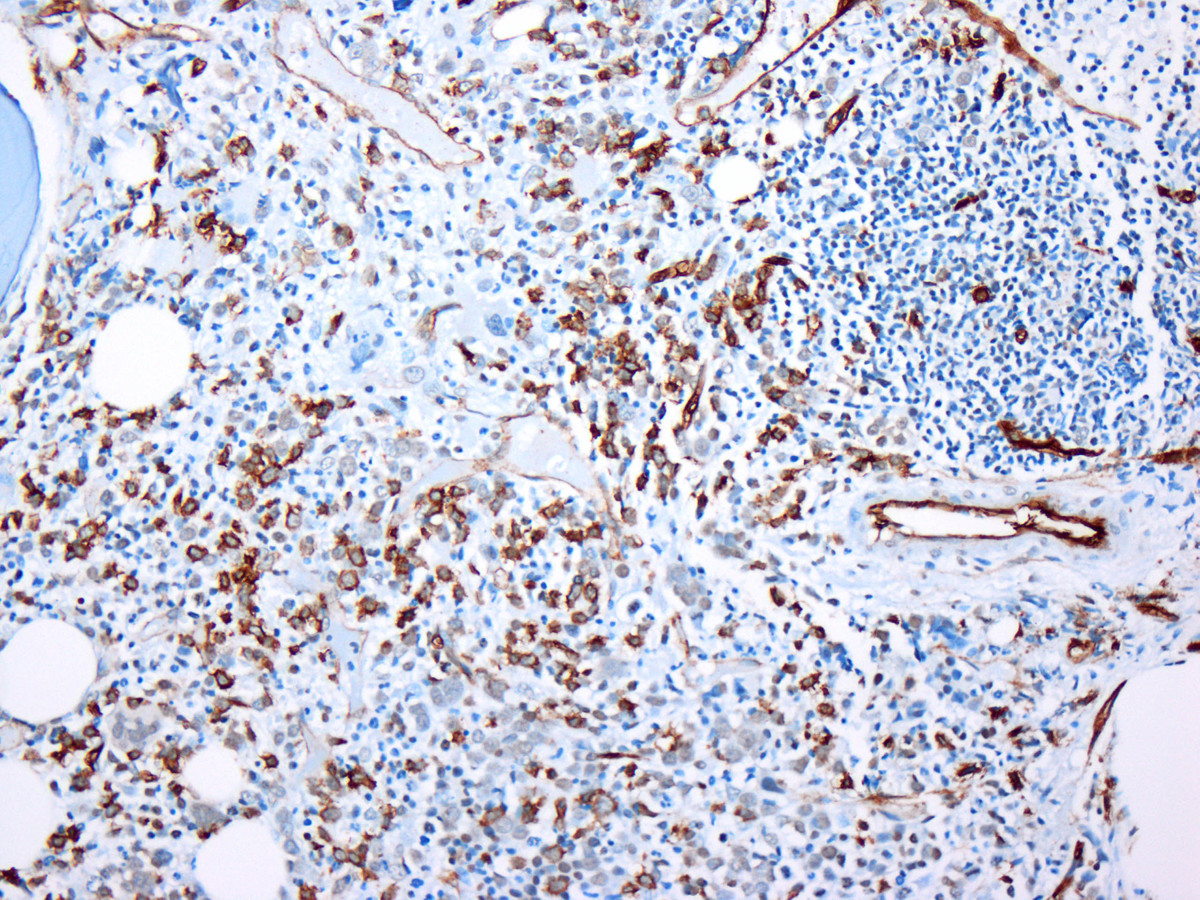 |
| Figure 4. Initial bone marrow biopsy: Immunohistochemistry for CD61 highlights dysplastic megakaryocytes. | 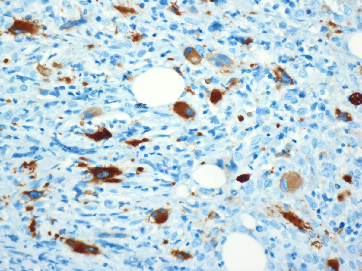 |
| Figure 5. Final bone marrow biopsy showing progression to pure erythroid leukemia: The markedly hypercellular bone marrow contains sheets of large blasts, as well as persistent CLL aggregates and dysplastic megakaryocytes (H&E). | 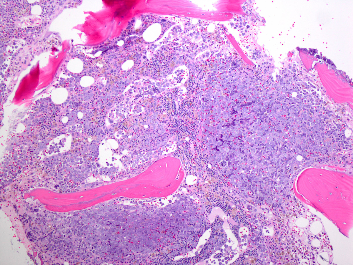 |
| Figure 6. Final bone marrow biopsy showing progression to pure erythroid leukemia: The blasts contain vesicular chromatin, multiple nucleoli, and smooth, amphophilic chromatin, consistent with erythroblasts (H&E). | 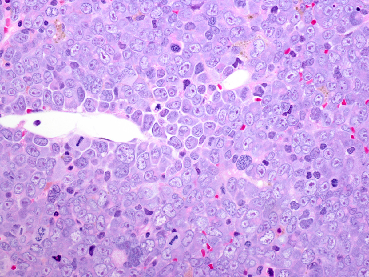 |
| Figure 7. Final bone marrow biopsy showing progression to pure erythroid leukemia: By immunohistochemistry, the blasts are glycophorin positive, confirming erythroid lineage. | 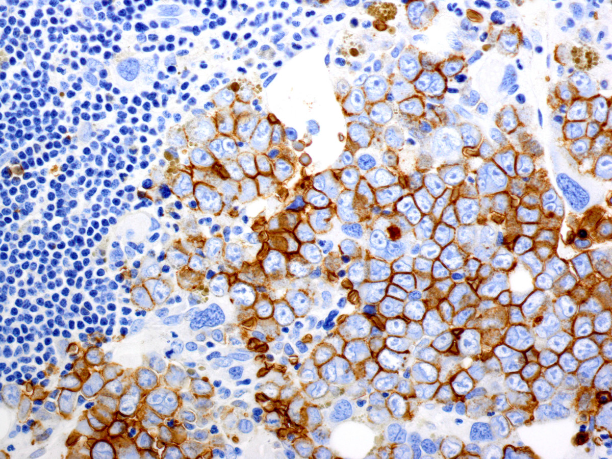 |
| Figure 8. Final bone marrow biopsy showing progression to pure erythroid leukemia: The background small lymphocytes are PAX5 positive, consistent with CLL. | 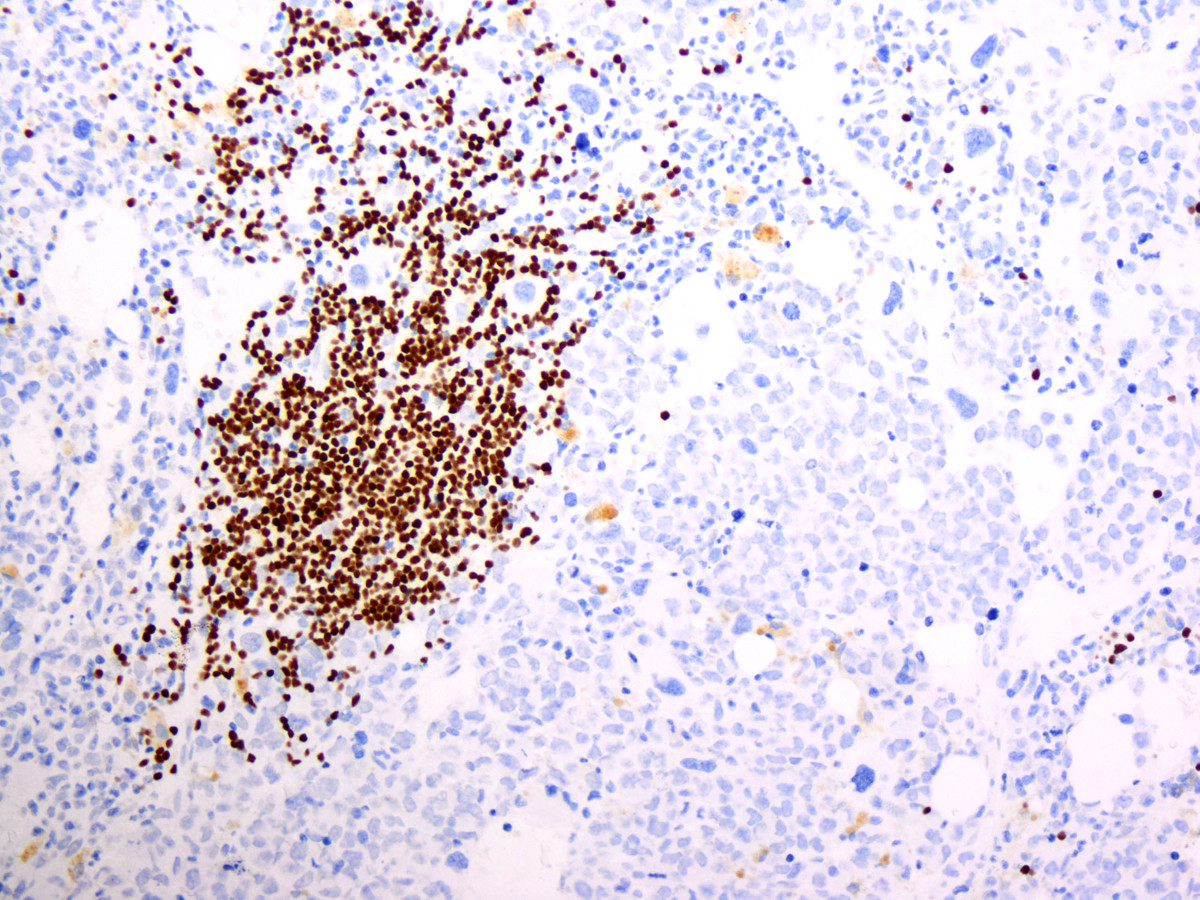 |
| Figure 9. Final bone marrow biopsy showing progression to pure erythroid leukemia: The bone marrow aspirate contains numerous large erythroid blasts with deeply basophilic cytoplasm, frequent cytoplasmic vacuoles, and often irregular and bizarre nuclei (Wright-Giemsa). |  |