Institution: Department of Hematopathology, The University of Texas MD Anderson Cancer Center, Houston
Additional authors:Yi Zhou, MD, PhD, Rajesh R Singh, PhD, Raja Luthra, PhD, Sergej N Konoplev MD
Session: T Lymphoblastic Leukemia/Lymphoma
HISTORY
42-year-old African American male in usual state of health until developing progressive cervical lymphadenopathy for approximately three months prior to initial presentation. The patient was presumptively diagnosed with Mumps then parotiditis by local physicians until undergoing left anterior cervical lymph node biopsy on 01/14/10. Pathological findings were consistent with T-lymphoblastic leukemia/lymphoma with expression of CD2, cytoplasmic CD3, surface CD3, dim CD5, CD7 and TdT (negative for CD1a, CD4, CD8). The patient was initially evaluated by the local oncologist on 01/26/10. Further staging evaluations such as PET/CT scan and bone marrow aspiration/biopsy were not performed in anticipation of undergoing these assessments at M. D. Anderson Cancer Center. The patient had not yet initiated therapy
DETAILS
Bone marrow smears, and formalin-fixed paraffin-embedded bone marrow biopsy and clot sections. • Peripheral blood smear demonstrates leukocytosis (15.8 K/UL) due to markedly increased blasts (58%). The blasts are of medium size, with irregular shaped nuclei, open nuclear chromatin, and moderate amount of light basophilic cytoplasm without granules. Occasional blasts demonstrate one or two small nucleoli. No Auer rods are identified. Granulocytes show complete maturation. Platelets are mildly decreased. Red blood cells are unremarkable. • Bone marrow differential count based on 500 cells revealed 75% blasts, 0% promyelocytes, 9% granulocytes, 1% eosinophils, 1% basophils 1% monocytes, 4% lymphocytes, 10% erythroid precursors. • Bone marrow aspirate smears and touch imprint are adequate for assessment. Tri-lineage hematopoiesis are relatively decreased. Blasts were markedly increased. Blasts are medium in size, with open nuclear chromatin, irregular shaped nuclei, and moderate amount of light basophilic cytoplasm with no granules. Occasional blasts demonstrate one or two medium sized nucleoli. Occasional blasts have rare small cytoplasmic vacuoles. No Auer rods are identified. Bone marrow biopsy and clot section showed cellularity of 80-90%. Megakaryocytes were present. Sheets of immature cells of medium size with open nuclear chromatin and moderate amount of cytoplasm are identified.
IMMUNOHISTOCHEMISTRY AND FLOW CYTOMETRY
We performed flow cytometry immunophenotypic studies using the bone marrow aspirate material of the specimen. The neoplastic cells are positive for CD2, CD3 (cytoplasmic and surface), CD5, CD7, CD19 (partial dim), CD34 (partial dim), CD38, CD58, CD81, and TdT. The neoplastic cells are negative for CD1a, CD4, CD8, CD10, CD13, CD14, CD15, CD20, CD22, CD41, CD56, CD64, CD66c, CD117, HLA-DR, myeloperoxidase, immunoglobulin kappa light chain immunoglobulin lambda light chain, and IgM. We performed immunohistochemical stain using fixed, paraffin embedded tissue of clot section. The neoplastic cells are negative for CD19, CD22, and CD79a.
CYTOGENETIC FINDINGS
50,XY,add(1)(q25),add(4)(q31),+8,+10,+13,+19[10] / 46,XY[10]
MOLECULAR FINDINGS
IgH PCR: No monoclonal immunoglobulin heavy chain gene rearrangements are detected by PCR analysis (polyclonal IgH amplification pattern). TCR-gamma PCR: Monoclonal T-cell receptor gamma chain gene rearrangements are detected by PCR analysis. Clonal TCR-gamma amplicon(s) utilize the V-gamma-I primer(s). TCR-beta PCR: Monoclonal T-cell receptor beta chain gene rearrangements are detected by PCR analysis. DNMT3A mutation Positive; R882C CGC->TGC
INTERESTING FEATURES
This case of T-lymphoblastic leukemia carried a mutation (R882C) in DNMT3A, DNA (cytosine-5)-methyltransferase 3A. DNMT3A is crucial for the methylation of unmodified DNA in CpG islands by converting cytosine to 5-methylcytosine. DNMT3A mediated DNA methylation is associated with gene silencing. Mutated DNMT3A has reduced enzymatic activity and attributes to the epigenetic deregulation increasingly recognized in acute myeloid leukemia (AML). Limited study has shown DNMT3A mutation is also present in T-ALL and associated with poor clinical outcome (GENES, CHROMOSOMES & CANCER 52:410–422 (2013) ).
PROPOSED DIAGNOSIS
T-lymphoblastic leukemia with DNMT3A mutation.
CONSENSUS DIAGNOSIS
T-acute lymphoblastic leukemia (T-IV; surface CD3-positive) with DNMT3A mutation
| Aspirate smear and trephine biopsy | 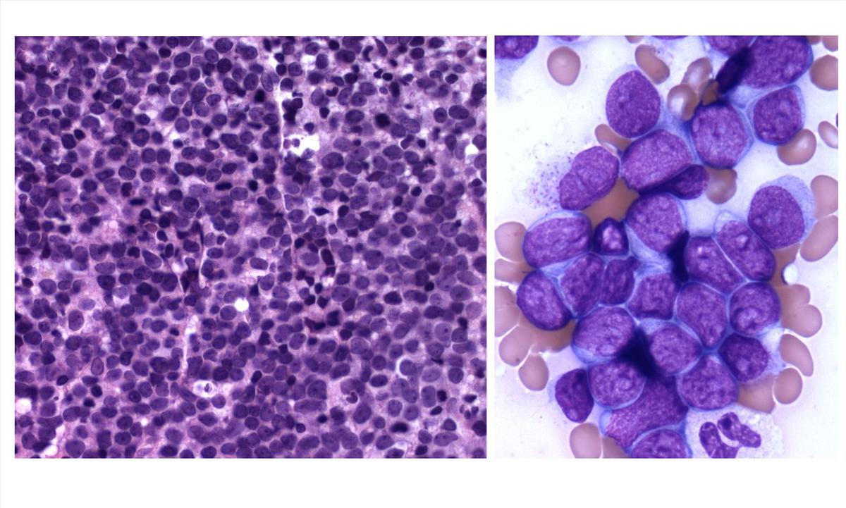 |
| Immunohistochemical stains |  |
| Flow cytometric analysis 1 | 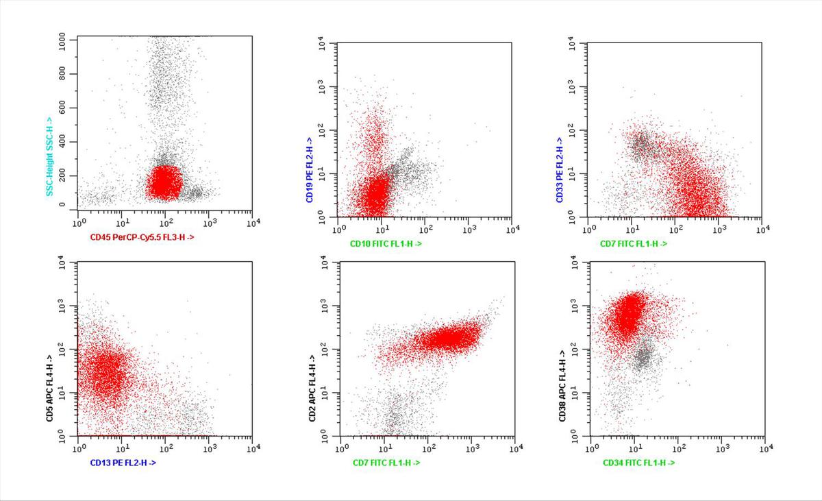 |
| Flow cytometric analysis 2 |  |
| DNMT3A mutation |  |