Institution: Department of Laboratory Medicine and Pathology, University of Minnesota
Additional authors:Michael A. Linden MD PhD, Robert W. McKenna MD.
Session: T Lymphoblastic Leukemia/Lymphoma
HISTORY
Patient is a 58 year old male with chief complaint of generalized weakness, fatigue and a history of unexplained anemia for about 2 months. CBC performed at that time revealed hemoglobin of 8.7 g/dL, WBC of 1.8x 10⁹/L with absolute neutrophil count of 0.2x 10⁹/L, platelets of 187x 109/L, and many circulating blasts. A bone marrow biopsy performed at an outside institution on 08/21/2012 was diagnosed as acute myeloid leukemia with T-cell antigens. Physical exam and CT of chest/Abd/Pelvis revealed no mass. The patient was transferred care to UMMC without further treatment.
DETAILS
Bone marrow smears, and formalin-fixed paraffin-embedded bone marrow biopsy and clot sections.
• The red blood cells appear normochromic. Poikilocytosis is minimal and nonspecific. Absolute neutropenia and lymphocytopenia are present. The platelets are slightly decreased with unremarkable morphology. Blasts are markedly increased (46%). • Bone marrow differential counts are: blasts 83%, granulocytes 8%, erythroid precursors 4%, monocytes 2%, eosinophils 1%, lymphocytes 2%• Bone marrow aspirate smears revealed complete neutrophilic and erythroid maturation. Megakaryocytes are present. Two different blast populations are identified. One population has large size, large nuclei, one to two prominent nucleoli and small amount of pale blue cytoplasm. The nuclei of these larger blasts show frequent nuclear grooves and notches. The second population has blasts with very little cytoplasm, rounded nucleus, smudged chromatin and smaller size compared to the other population.Trephine and clot sections show increased marrow cellularity, 85-90%.Megakaryocytes are present but decreased in numbers. Sheets and clusters of blasts occupy at least 80% of the cellular composition of the bone marrow.IMMUNOHISTOCHEMISTRY AND FLOW CYTOMETRY
Bone marrow aspirate was analyzed on flow cytometry. The blast gate (dim CD45 and low side scatter) accounts for greater than 95% of leukocytes (CD45+ cells). The blasts express CD2, partial CD7, partial CD13, partial CD15, partial CD34, CD38, dim CD45, CD58, dimCD117, partial HLA-DR, partial cytoplasimic CD3 and nuclear TdT, but lack CD1a, surface CD3, CD4, CD5, CD8, CD10, CD14, CD19, CD20, CD22, CD33, CD36(dim to absent), CD56, CD61, glycophorin A and cytoplasmic CD79a and cytoplasmic myeloperoxidase.
CYTOGENETIC FINDINGS
47, XY, +4 [14]/ 46, XY [6]
INTERESTING FEATURES
The leukemic blasts in this case are positive for T-cell antigen, CD2, cytoplasmic CD3, CD7; myeloid antigen CD13, CD15, CD117, and stem cell marker CD34, TdT, but negative for CD1a, CD4, CD5, and CD8. This immunophenotype is characteristic of the recently described early T-precursor (ETP) lymphoblastic leukemia/lymphoma.
ETP lymphoblastic leukemia is associated with very poor outcome in pediatric patients. This patient is 58 year old. His disease showed resistance to standard H-CVAD induction therapy. Approximately 10% blasts were still present at week 6 after cycle 1A and 1B treatment. Complete remission was achieved after salvage therapy with Nelarabine. The patient later received double cord-blood transplant and has been remaining in remission for 2 months.PROPOSED DIAGNOSIS
Early T-precursor lymphoblastic leukemia
CONSENSUS GROUP: ADDITIONAL INFORMATION/STUDIES
Additional immunostains performed by the conference consensus group:
CD3: Positive
CONSENSUS DIAGNOSIS
T-acute lymphoblastic leukemia, early T-precursor immunophenotype
| Biopsy and aspirate at diagnosis | 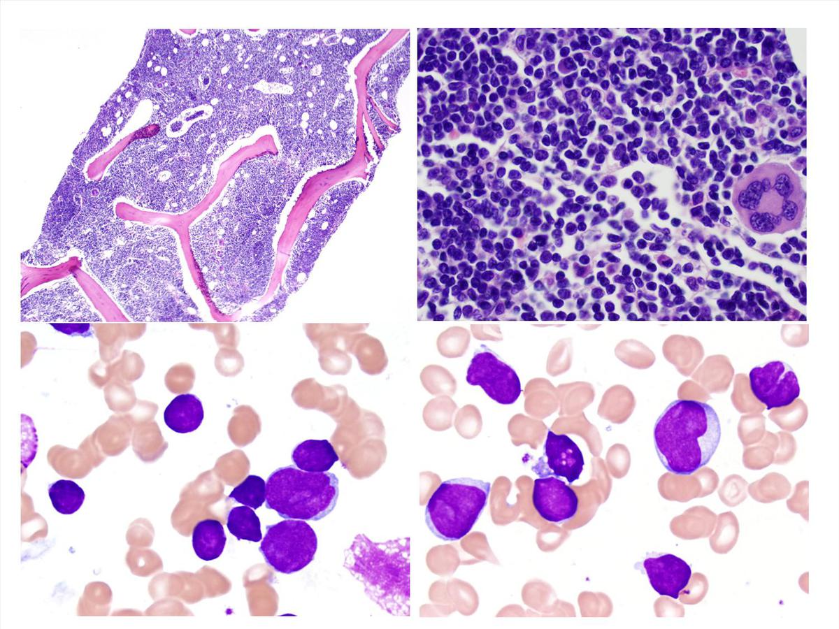 |
| flow cytometry immunophenotype at diagnosis 1 |  |
| flow cytometry immunophenotype at diagnosis 2 | 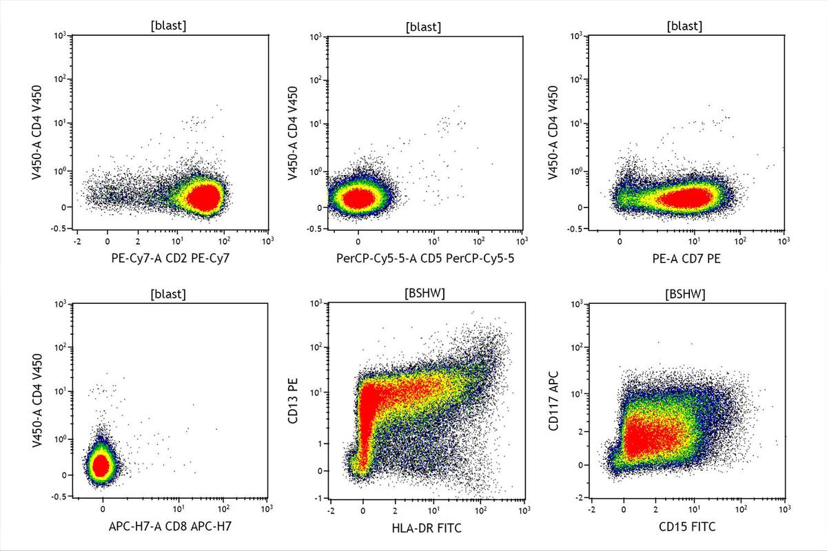 |
| Biopsy and aspirate at day 42 |  |
| flow cytometry immunophenotype at day 42 1 | 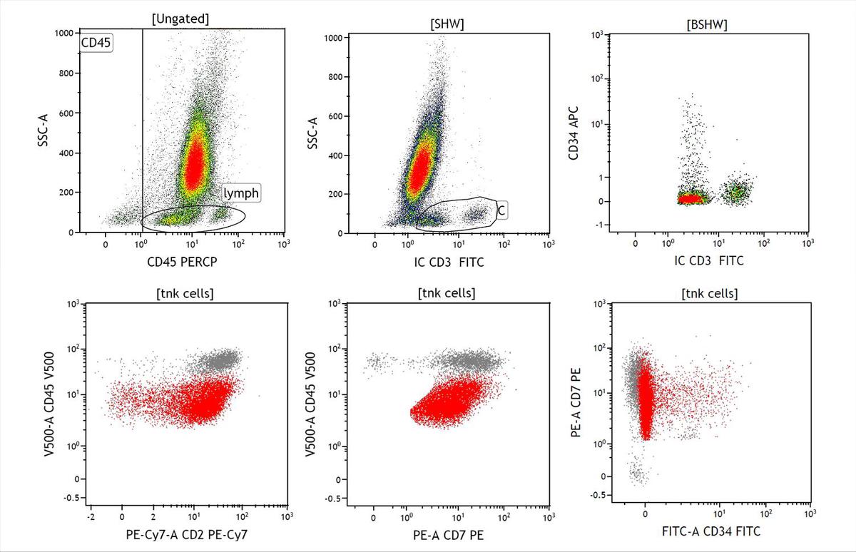 |
| flow cytometry immunophenotype at day 42 2 | 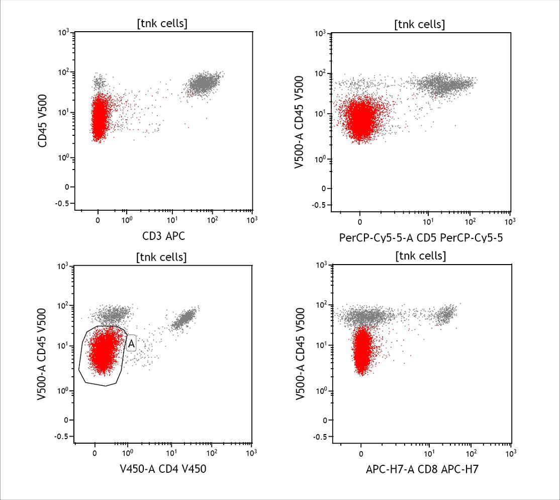 |
| CD3 IHC (consensus group additional stain) |  |