Institution: New York-Presbyterian Hospital/Weill Cornell Medical Center
Additional authors:Wayne Tam MD, PhD, Attilio Orazi MD
Session: AML with myelodysplasia-related changes
HISTORY
45 year old woman in good health until November 2012 when she developed progressive fatigue. Subsequently developed headache and mild dyspnea on exertion. Seen by internist on 11/12/2012 and CBC showed pancytopenia with blasts in peripheral blood.
DETAILS
The marrow is fibrotic and shows increased cellularity (~60-70%). M:E ratio appears decreased due to decreased granulopoiesis and slightly increased erythropoiesis. Granulopoiesis demonstrates complete maturation. Blasts are scattered and slightly increased (see also results of flow cytometry and immunohistochemistry). Erythropoiesis demonstrates complete normoblastic maturation. Megakaryocytes are markedly increased and atypical. They are predominantly small with hypolobated, hyperchromatic nuclei. Micro megakaryocytes are identified. In addition, an increased number of small lymphocytes and mature plasma cells are scattered throughout the marrow. Clot section is inadequate. It consists only of aparticulated blood clots. Reticulin stain shows marked (3+) reticulin fibrosis.
IMMUNOHISTOCHEMISTRY AND FLOW CYTOMETRY
Immunohistochemistry:
CD34 5-10%
CD117 10%
MPO 20%
Glycophorin C 30%
CD42b Markedly increased megakaryocytes with atypia
INTERPRETATION: Immunoperoxidase staining was performed on the bone marrow biopsy to evaluate the number of blasts and to assess the marrow cell composition. CD34 and CD117 stain slightly increased scattered blasts. CD117 is also positive in scattered mast cells and erythroid precursors. MPO demonstrates decreased granulopoiesis. Glycophorin C stains maturing erythropoiesis. CD42b shows numerous dysplastic megakaryocytes, including micromegakaryocytes.
Flow cytometry:
Analysis of the faint CD45 cell population shows increased myeloblasts, accounting for 10% of total cells analyzed. These blasts are CD34+, CD117+, HLA-DR (small subset), CD13+, CD33+, and CD7+. These flow cytometry findings support the presence of excess blasts.
CYTOGENETIC FINDINGS
44,XX,-3,der(5)t(3;5)(p13;q?22),-7,-12,-20,+mar1,+mar2[6]/46,XX[7]
An abnormal karyotype with monosomy of chromosomes 3, 7, 12 and 20, an unbalanced der(5)t(3;5) translocation resulting in partial loss of 5q, and two marker chromosomes was observed in six of 13 evaluated metaphase cells. A normal female karyotype was observed in the remaining seven metaphase cells.
MOLECULAR FINDINGS
INTERESTING FEATURES
The overall findings support a diagnosis of RAEB-2 with fibrosis. Acute panmyelosis with myelofibrosis (APMF) could also be considered in the differential diagnosis. However, the relatively low percentages of blasts (5% in the peripheral blood; 8-10% in the bone marrow) associated with the presence of decreased granulopoiesis (i.e., lack of panmyelosis) argues against this alternative diagnostic possibility. Although RAEB-2 with fibrosis and APMF have many overlapping features (e.g., similar degree of marrow fibrosis, multilineage dysplasia, and often complex karyotype which include anomalies of chromosome 5 and 7), by carefully applying an integrated diagnostic approach, separation can be achieved in most cases.
PROPOSED DIAGNOSIS
Myelodysplastic syndrome, refractory anemia with excess blasts-2 (RAEB-2), with fibrosis, in bone marrow.
CONSENSUS DIAGNOSIS
Myelodysplastic syndrome, refractory anemia with excess blasts-2 (RAEB-2), with fibrosis
| The marrow shows increased cellularity (~60-70%). | 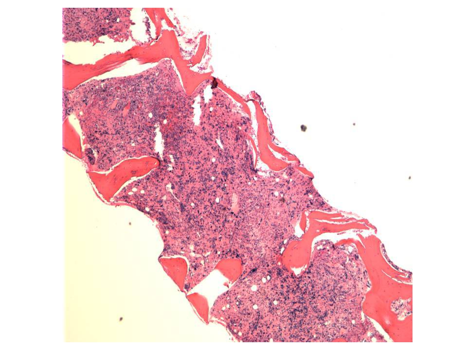 |
| Megakaryocytes are markedly increased. | 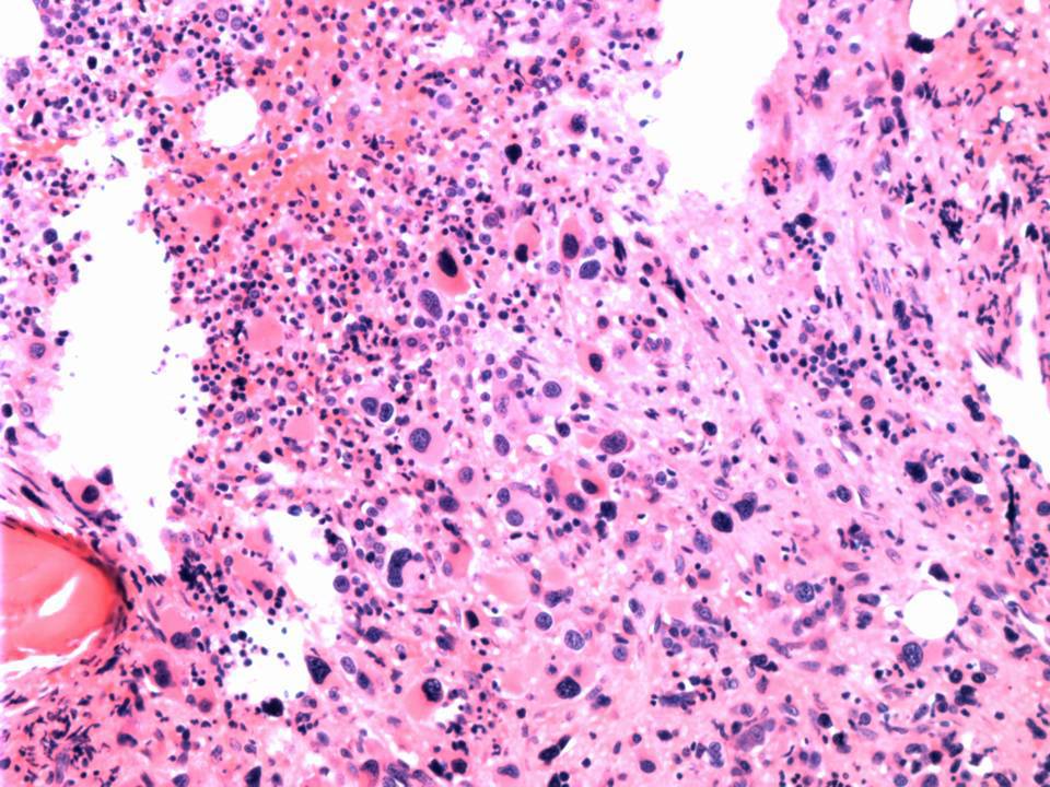 |
| M:E ratio appears decreased due to decreased granulopoiesis and slightly increased erythropoiesis. Granulopoiesis demonstrates complete maturation. Erythropoiesis demonstrates complete normoblastic maturation. | 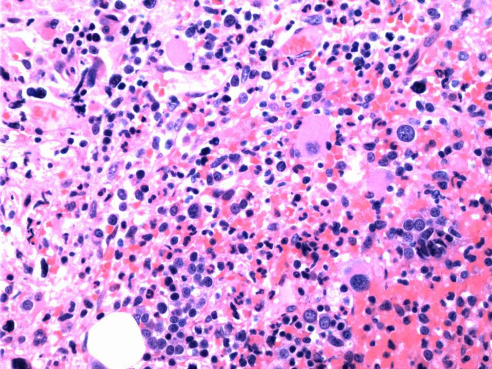 |
| Megakaryocytes are markedly increased and atypical. | 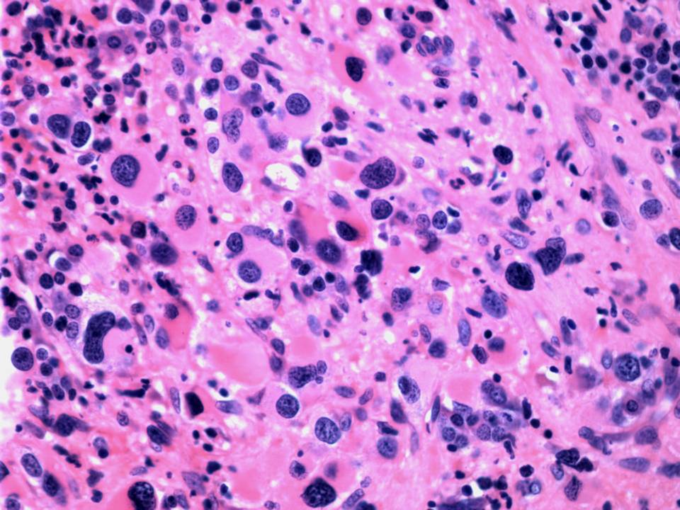 |
| Reticulin stain shows marked (3+) reticulin fibrosis. | 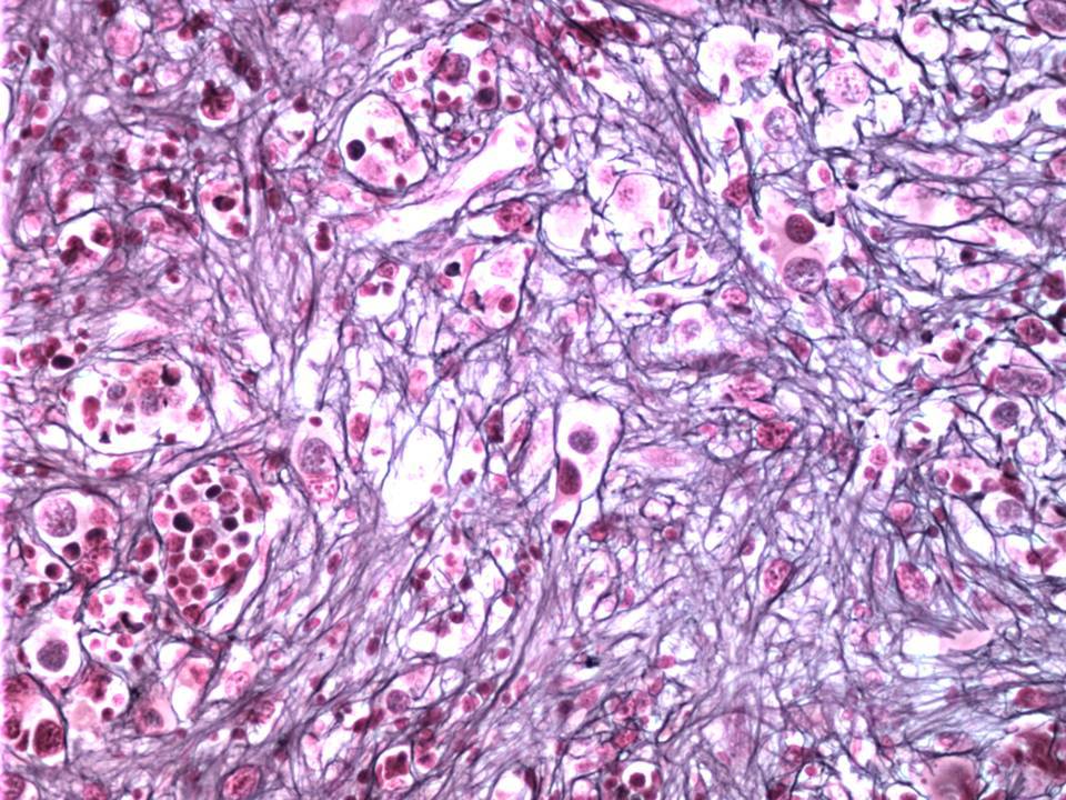 |
| CD34 stains slightly increased scattered blasts. MPO demonstrates decreased granulopoiesis. Glycophorin C stains maturing erythropoiesis. CD42b shows numerous dysplastic megakaryocytes, including micromegakaryocytes. | 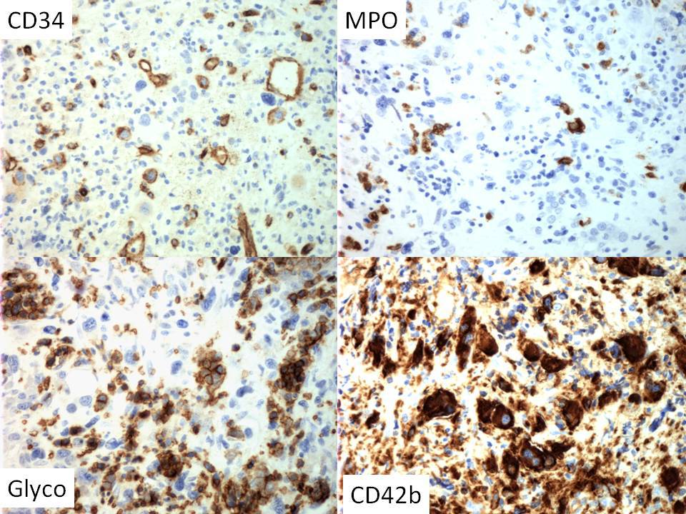 |