Institution: University of Arkansas for Medical Sciences
Additional authors:Jorge F Jimenez MD, National Park Medical Center
Session: Extramedullary manifestations of myeloid neoplasms
HISTORY
A 35 year old male with no significant past medical history had a sebaceous cyst excised from his left chest area in May 2012. The patient presented to his primary care physician in June 2012 with a painless golf ball sized cystic mass in the right axilla. The preoperative diagnosis was sebaceous cyst. Additional laboratory studies were not performed at the time of the biopsy.
DETAILS
Skin Biopsy:
The H and E stained sections of the formalin fized, paraffin-embedded skin showed a dermal infiltrate of intermediate sized cells with open chromatin and nucleoli. Occasional mitotic figures were noted. Hematopoietic cell lineage was not apparent based on morphologic features (see immunohistochemistry). Bone Marrow Aspirate and Biopsy (Wright stain aspirate):A subsequent bone marrow biopsy showed a hypercellular marrow for age involved by acute myeloid leukemia.IMMUNOHISTOCHEMISTRY AND FLOW CYTOMETRY
Skin biopsy, immunohistochemistry:
A panel of immunohistochemical stains was performed on formalin fixed paraffin embedded sections of the right armpit skin biopsy. The neoplastic cells were positive for CD45, CD43, CD117, TdT (subset), lysozme (subset), CD68 (subset), CD163 (subset), MPO (subset) and CD4. The tumor cells were negative for lymphoid markers (CD20, PAX-5 and CD3), CD56 and CD34. Flow cytometry (bone marrow aspirate): The corresponding flow cytometric analysis showed 77% blasts with monocytic differentiation and positive for HLA-DR, CD13, CD33, CD117, CD64 (subset), CD4 (subset), CD7 (subset), CD11b, (subset), CD11c (subset) and MPO (subset).CYTOGENETIC FINDINGS
Bone Marrow Aspirate:
Normal male karyotype: 46,XY[20].MOLECULAR FINDINGS
Skin Biopsy:
Molecular tests were not performed.Bone Marrow Aspirate:FISH analysis was negative for 11q23 translocations. Molecular studies were positive for FLT3 D835Y mutation and negative for FLT-3 ITD, NPM1 and CEBPA mutations.INTERESTING FEATURES
The diagnosis was unexpected given the lack of prior history and painless axillary mass. In this case, myeloid sarcoma was associated with concurrent acute myeloid leukemia with monocytic differentiation based on flow immunophenotype. Although the molecular genetics of the extramedullary tumor were not assessed, the bone marrow aspirate cells were positive for FLT3 D835Y mutation.
PROPOSED DIAGNOSIS
Myeloid sarcoma with concurrent acute myeloid leukemia.
CONSENSUS DIAGNOSIS
Acute myeloid leukemia with FLT3 D835Y mutation involving bone marrow and axillary skin mass (myeloid sarcoma)
| Skin Biopsy: H and E stain, 40x | 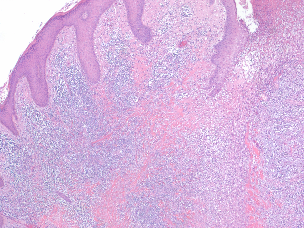 |
| Skin Biopsy: H and E stain 1000x |  |
| Skin Biopsy: CD45 immunostain 400x | 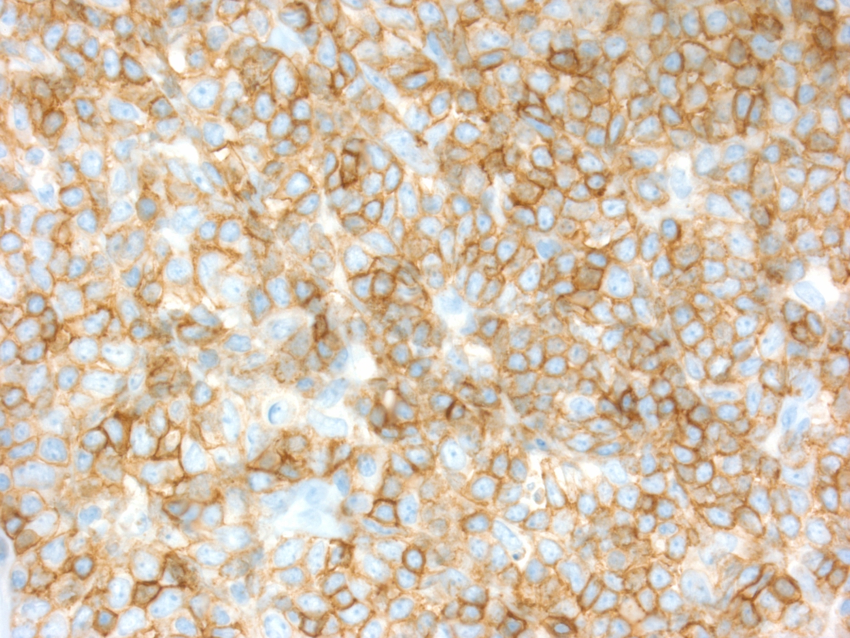 |
| Skin Biopsy: CD117 immunohistochemical stain, 400x |  |
| Skin Biopsy: CD68 Immunohistochemical stain, 400x |  |
| Skin Biopsy: TdT immunohistochemical stain, 400x | 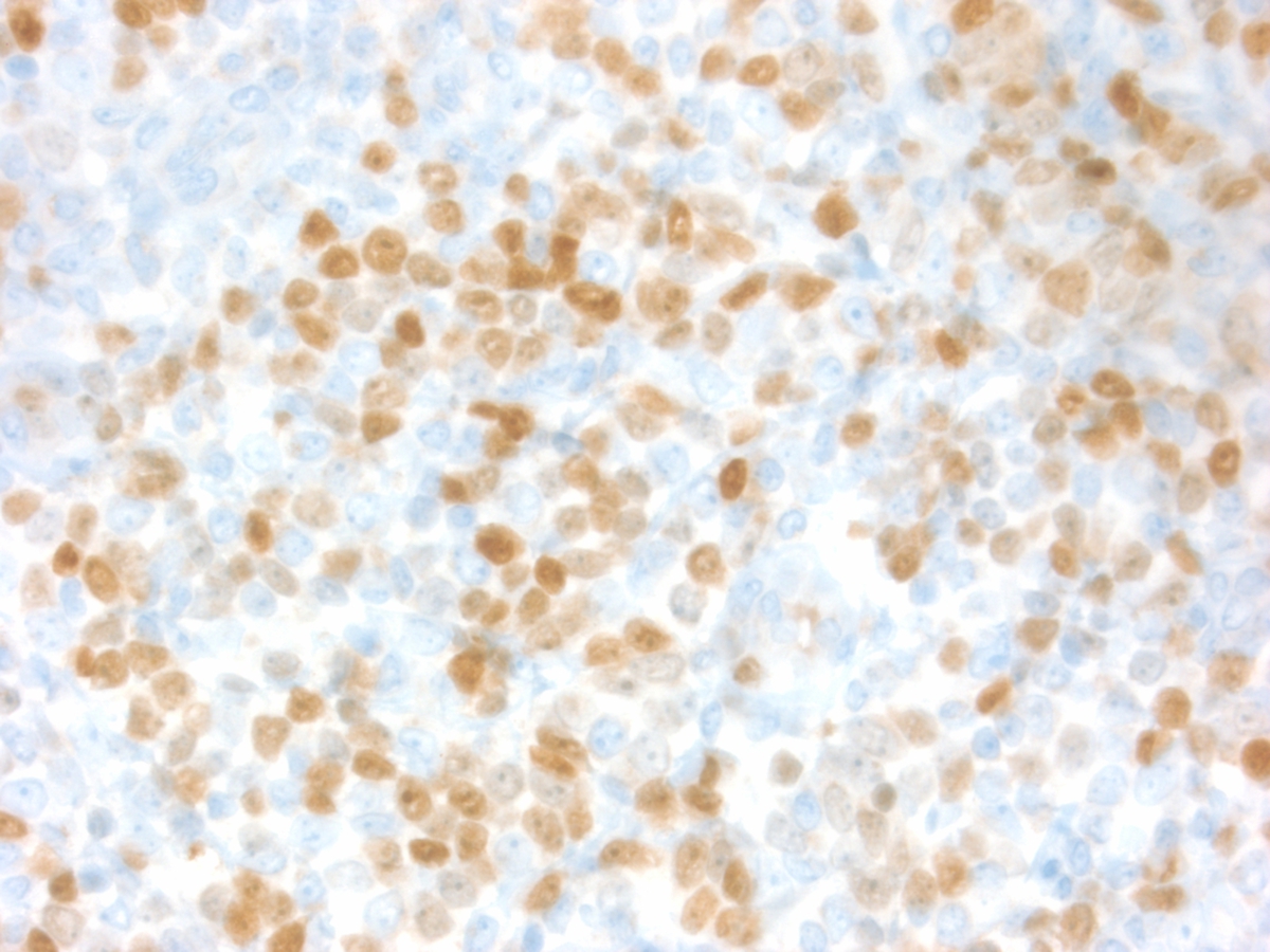 |
| Skin Biopsy: Myeloperoxidase immunohistochemical stain, 400x |  |
| Skin Biopsy: CD34 immunohistochemical stain, 400x | 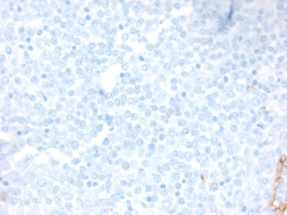 |
| Bone marrow aspirate, Wright stain, 1000x | 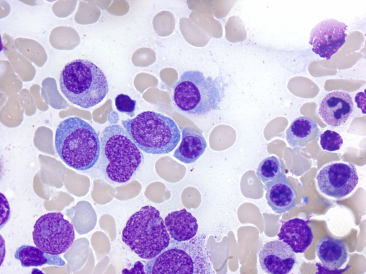 |