Institution: University of Arkansas for Medical Sciences
Session: Therapy-related myeloid neoplasms
HISTORY
A 56 year old female presented in February 2011 with complaints of confusion and generalized bone pain. Work up at the referring institution revealed hypercalcemia, hyperviscosity and IgG multiple myeloma (MM). She was referred to the Myeloma Institute where she was found to have obliteratve bone marrow plasmacytosis, mild anemia, thrombocytopenia and multiple MRI focal lesions with PET avidity. The patient was treated on total therapy 4 (TT4) protocol for newly diagnosed MM. The patient received one induction cycle with M-VDT-PACE (melphalan-bortezomib/dexamethasone/thalidomide- cisplatin/ adriamycin/cyclophophamide/etoposide) with stem cell collection in March 2011, two fractionated melphalan plus VDT with autologous stem cell transplants in May and July 2011, consolidation with VDT-PACE in August 2011 and started VRD maintenance (bortezomib, lenalidomide dexamethasome) in November 2011. The patient achieved stringent complete remission for MM.
A CBC in July 2012 revealed pancytopenia (Hgb = 9.9; ANC = 0.1; platelet count = 78 K) with absolute monocytosis including immature forms. Additional labs at this time revealed a normal LDH, B2M and albumin with mildly decreased total protein. Serum protein electrophoresis revealed trace M protein with possible IgG lambda band detected by serum immunofixation.DETAILS
Bone marrow aspirate (Wright stain) and core biopsy:
The bone marrow core biopsy was subcortical and insufficient for evaluation. The bone marrow aspirate smears showed erythroid predominance (M:E ratio 0.5:1) with left shifted and dyspoietic erythroid maturation (megaloblastoid, nuclear budding and multinucleation). Granulopoiesis was dyspoietic and left shifted myelomonocytic maturation and increased blasts.IMMUNOHISTOCHEMISTRY AND FLOW CYTOMETRY
Flow cytometric analysis performed on the aspirate cells showed 36% monocytes with expression of HLA-DR, CD14 (variable), CD15, CD13, CD33, CD4 and CD56.
CYTOGENETIC FINDINGS
Cytogenetics revealed 46,XX,t(4;11)(q21;q23)[16]/46,XX[4].
The t(4;11)(q21;q23) is associated with a fusion of the MLL gene and AFF1 gene.MOLECULAR FINDINGS
FISH analysis showed t(11q23) in 44% of analyzed nuclei.
bNo evidence of numeric or structural abnormalities of chromosomes 5, 7, 8, 13, or 20.INTERESTING FEATURES
The usual course of t-MN in MM patients treated on total therapy protocols is a prolonged MDS phase with progressive cytopenias, progressing to acute myeloid leukemia (AML) in a subset of patients with MDS chromosomal abnormalities (del20q, -7, del7q, -5, del5q and complex karyotype). This patient is unusual with a rapid onset of acute myeloid leukemia within 4 months of a morphologically unremarkable, albeit cytogenetically abnormal bone marrow evaluation, and the presence of a balanced translocation involving 11q23.
PROPOSED DIAGNOSIS
Therapy related myeloid neoplasm (t-AML)
CONSENSUS DIAGNOSIS
Therapy related myeloid neoplasm; acute myeloid leukemia with t(4;11)(q21;q23)
| Bone marrow aspirate (Wright stain): Erythroid dyspoeisis and increased blasts | 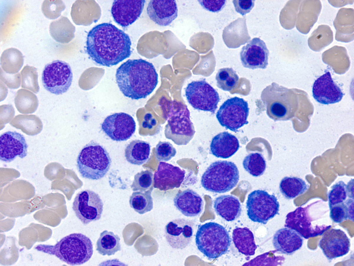 |
| Bone marrow aspirate (Wright stain); Monoblasts, 1000x | 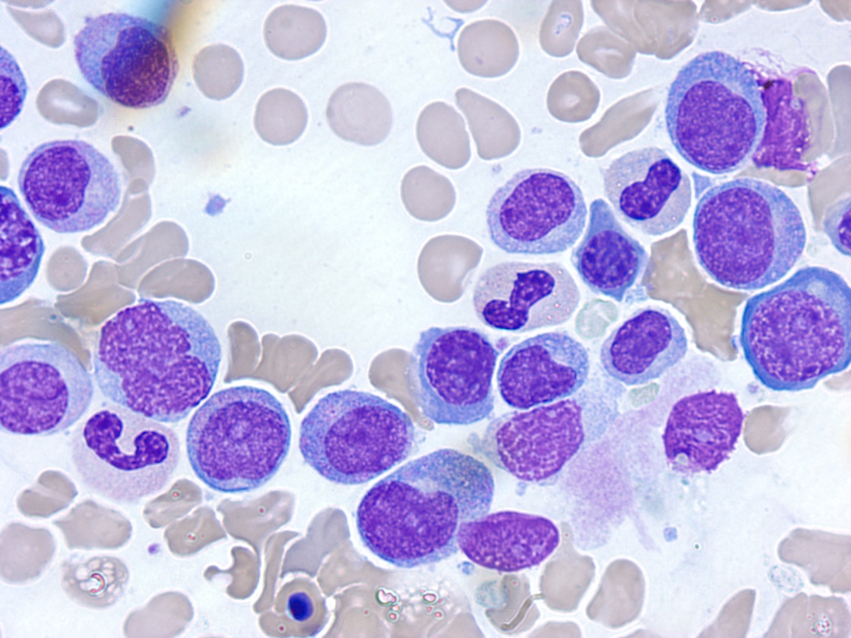 |
| Bone marrow aspirate flow phenotype | 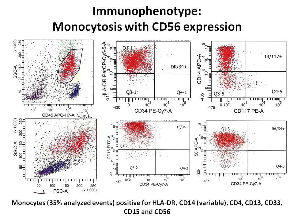 |