Institution: Mayo Clinic
Additional authors:Rong He, M.D., Patricia T. Greipp, D.O.
Session: Therapy-related myeloid neoplasms
HISTORY
The patient is a 76 year-old female with an original diagnosis in 1985 of monoclonal gammopathy of undetermined significance (MGUS) with an IgG kappa serum monoclonal protein of 1.8 g/dL. By 1994, the serum M protein had reached 3.8 g/dL and bone marrow biopsy showed multiple myeloma with 30-40% plasma cell marrow involvement. Through 2008 she was treated with a combination of vincristine, carmustine, melphalan, cyclophosphamide, prednisone, and alpha interferon. At that time her myeloma had gradually progressed and serum M protein was 1.5 g/dL and 20% bone marrow biopsy plasma cells. Lenalidomide with dexamethasone was begun and serum M protein levels again decreased and then remained relatively stable; however, by 2010 she gradually developed cytopenias.
By 2012, the cytopenias continued with hemoglobin less than 10.0 g/dL, absolute neutrophil count of 0.6x10^9 /L, platelets of 66x10^9 /L, and lenalidomide was discontinued. A bone marrow biopsy was then performed; the findings are the subject of the following discussion.DETAILS
Peripheral blood smears were prepared with Wright Giemsa stain. Iliac crest bone marrow aspirate and biopsy were taken; the aspirate was prepared with Wright Giemsa stain and the biopsy was stored in B5-formalin mixed solution until paraffin embedding and histologic sections stained with hematoxylin and eosin. Additional studies included flow cytometry studies, butyrate esterase/chloroacetate esterase cytochemical stains, immunohistochemistry, cytogenetic analysis, and fluorescence in situ hybridization (FISH).
Morphologic review demonstrated increased (20%) plasma cells, which were distributed in small aggregates and cytologically were atypical with large nucleoli. Blasts were borderline increased (5%) and demonstrated medium-sized nuclei, fine chromatin, and prominent nucleoli.IMMUNOHISTOCHEMISTRY AND FLOW CYTOMETRY
Immunohistochemistry was performed on the sections of the bone marrow biopsy with antibodies to CD3, CD20, CD34, CD61, and CD138. Notably, the atypical plasma cells were positive for CD138, and scattered blasts were positive for CD34. Flow cytometric studies demonstrated kappa light chain restriction amongst the neoplastic plasma cells.
CYTOGENETIC FINDINGS
Classic chromosome G-banding studies were performed; of 20 metaphases, 13 were normal and 7 had t(3;12;21)(q26.2;q24.3;q22). Dual color double fusion fluorescence in situ hybridization (DFISH) revealed t(3;21)(MECOM/RUNX1) in 7.8% of nuclei, consistent with the above result in the chromosome studies.
MOLECULAR FINDINGS
Not performed.
INTERESTING FEATURES
This case demonstrates therapy related myelodysplasia in the setting of a background plasma cell proliferative disorder; specifically, a three-way translocation in which two involved partners were MECOM and RUNX1.
PROPOSED DIAGNOSIS
1) Plasma cell myeloma
2) Myelodysplastic syndrome, refractory anemia with excess blasts-1CONSENSUS DIAGNOSIS
1) Therapy related myeloid neoplasm, myelodysplastic syndrome, with t(3;12;21) [5% blasts]
2) Plasma cell myeloma| Bone marrow aspirate | 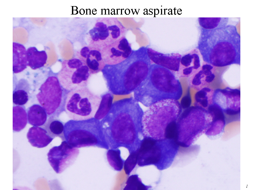 |
| Bone marrow aspirate | 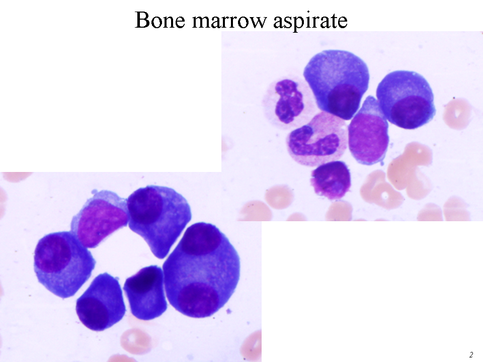 |
| Bone marrow aspirate | 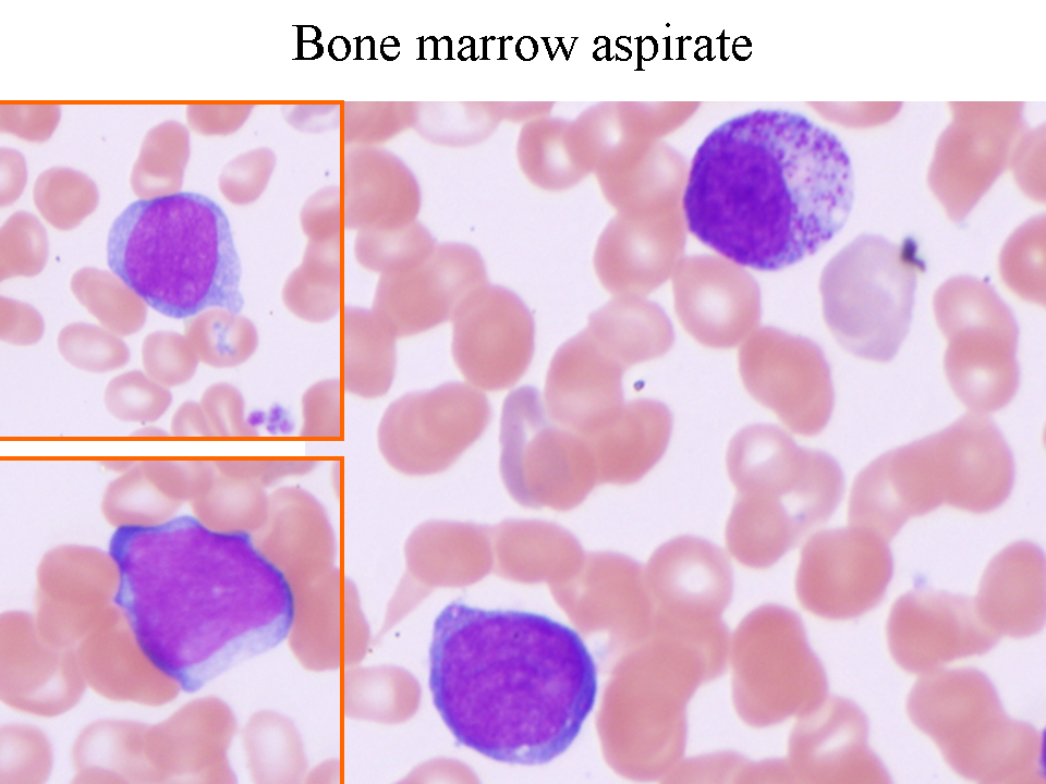 |
| Bone marrow biopsy section | 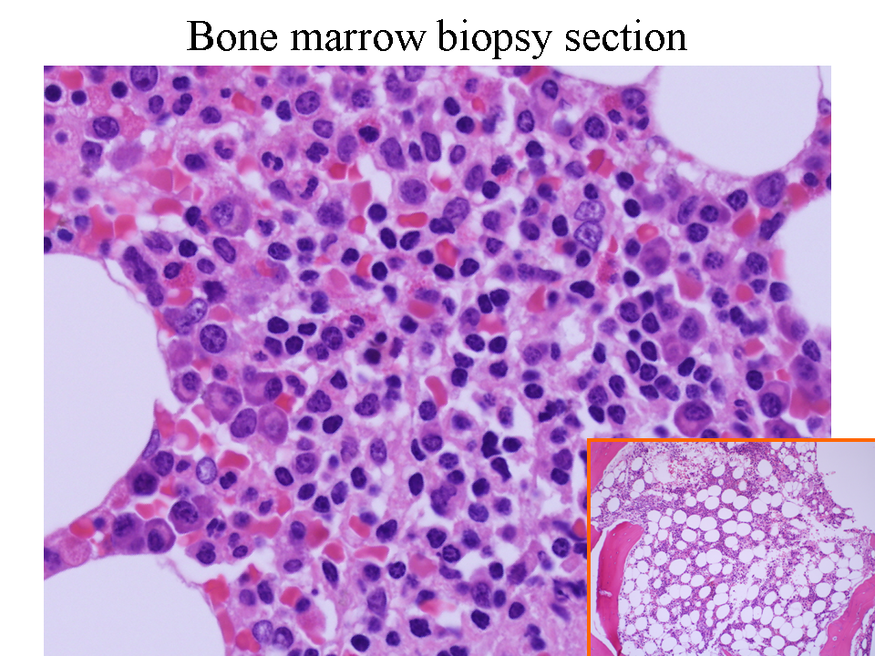 |
| Bone marrow biopsy section (CD34) | 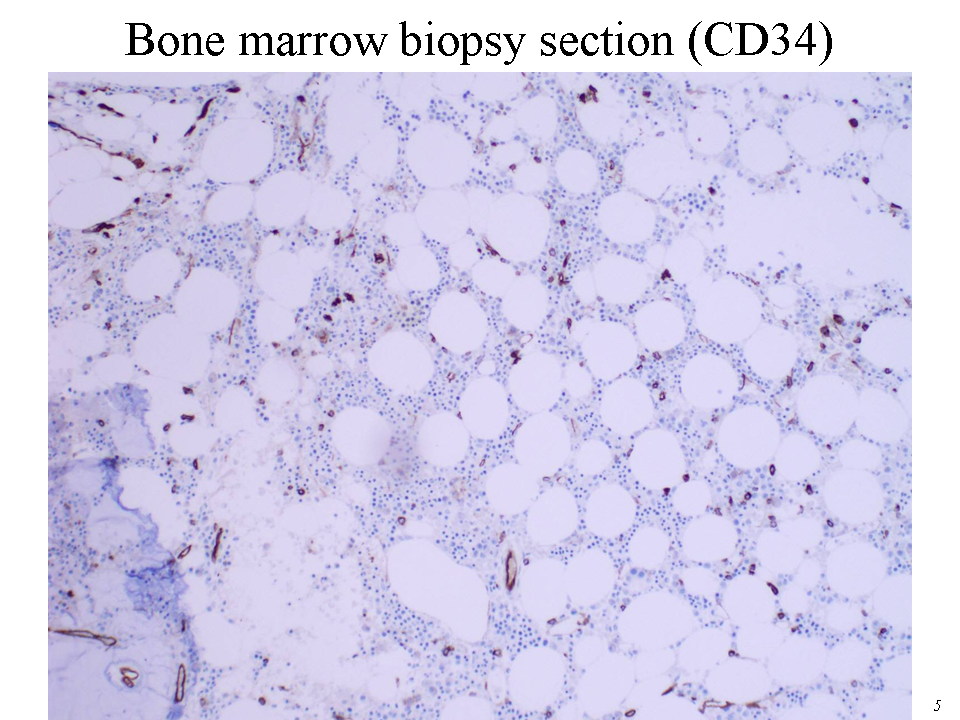 |
| Bone marrow biopsy section (CD34) | 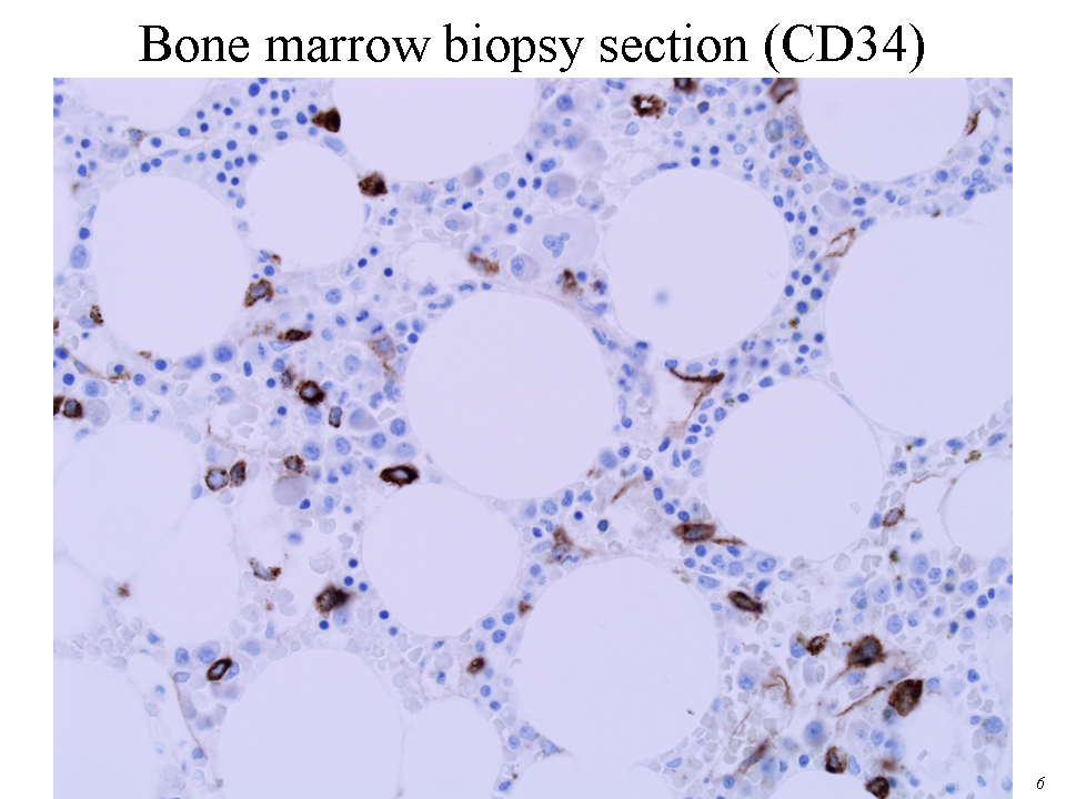 |
| Bone marrow biopsy section (CD138) | 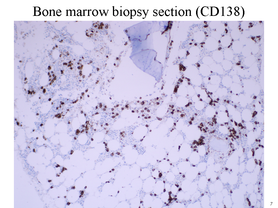 |
| Metaphase G-banded karyogram (Courtesy of Patricia T. Greipp, D.O.) | 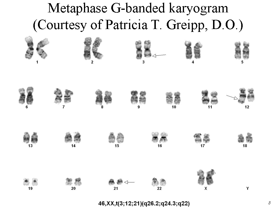 |
| Metaphase G-banded karyogram (Courtesy of Patricia T. Greipp, D.O.) | 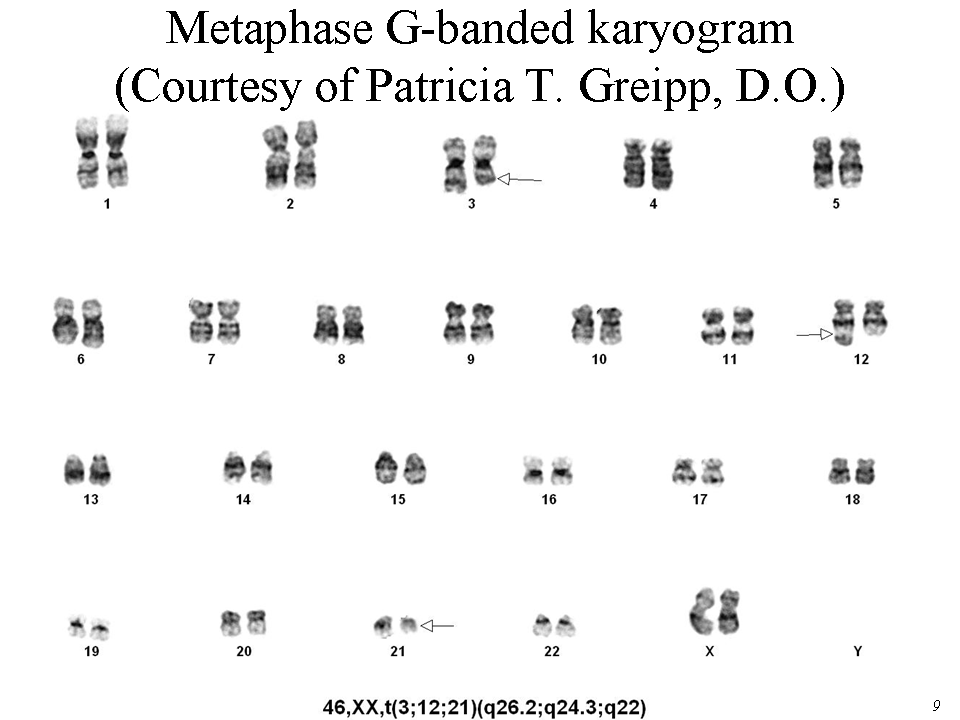 |
| Bone marrow aspirate FISH (Courtesy of Patricia T. Greipp, D.O.) | 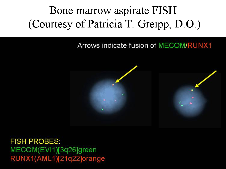 |