Institution: University of Wisconsin School of Medicine and Public Health
Session: Extramedullary manifestations of myeloid neoplasms
HISTORY
A 39 year old male presented to his primary physician with redness and swelling around a scratch on his right shin. His past history was significant for idiopathic dilated cardiomyopathy treated with an indwelling defibrillator and ablations for atrial fibrillation. His CBC was normal. He was diagnosed with cellulitis and treated with antibiotics, but the skin lesion became larger and partially necrotic. Over the course of a week, concern for necrotizing fasciitis resulted in three surgical debridements. Biopsy at the outside hospital (and subsequent debridement specimens) showed a neutrophilic infiltrate without vasculitis and without microorganisms. Combined with the negative cultures and organism stains, this raised concern for Sweet's syndrome vs. pyoderma gangrenosum. At the outside hospital, his CBC showed WBC - 7.1 K/ul with normal differential, Hgb-13.8 mg/dL, PLT = 200K/ul, and MCV = 102. On the 6th day of hospitalization it was WBC = 10.9 K/ul with 1% blasts, 3% immature myeloids, Hgb = 10.8 mg/dL, and platelets = 115. The next day he was started on prednisone to treat the leg lesion, but became hypotensive, requiring pressors, and was transferred to our institution (blood cultures were negative). On admission his CBC showed WBC = 22.4 K/uL with 1344 blasts, Hgb 9.5 mg/dL, and PLT = 144 K/ul. Peripheral smear showed dysplastic, hypogranulated neutrophils and bands as well as circulating blasts. Marrow biopsy showed similar findings with 24% blasts. The patient's leg ulcer was very large, but non-purulent (see image). Induction chemotherapy was instituted which showed improved marrow findings but residual blasts (6-12%) at days 10 and 17 with residual cytogenetic abnormalities. For insurance reasons, the patient was transferred to another hospital with a plan for re-induction and eventual skin grafting.
DETAILS
Iliac crest marrow biopsy with formaldehyde fixation and decalcification and H/E staining. Aspirate and peripheral smear stained with Wright's stain. The marrow showed 24% myeloblasts and 11% promyelocytes. Myeloid cells were dysplastic with hypogranulation and nuclear-cytoplasmic dyssynchrony. Megakaryocytes showed occasional cells with separate nuclear lobes and rare erythroids showed nuclear irregularities. The degree of dysplasia combined with the very acute presentation suggested an inv3 or t(6;9) cytogenetic abnormality. The outside skin biopsy has been requested for review (described above).
IMMUNOHISTOCHEMISTRY AND FLOW CYTOMETRY
Blasts positive for CD13 and CD117, with dim CD33 and heterogeneous expression of CD11b, CD15, CD16, CD34, CD64, HLA-DR. Negative for MPO, TdT, and other lymphoid markers.
CYTOGENETIC FINDINGS
Positive for t(6;9)(p23;q34); DEK-NUP24 translocation.
MOLECULAR FINDINGS
INTERESTING FEATURES
A very acute onset with essentially normal CBC presenting with pyoderma gangrenosum which was misinterpreted as necrotizing fasciitis and debrided surgically multiple times before steroid therapy was instituted. Also a nice example of the dysplasia in t(6;9) AML which should not be labeled AML with myelodysplastic changes.
PROPOSED DIAGNOSIS
Acute myeloid leukemia with t(6;9)(p23;q34); DEK-NUP24 translocation with concomitant pyoderma gangrenosum.
CONSENSUS DIAGNOSIS
Acute myeloid leukemia with t(6;9)(p23;q34); DEK-NUP24 translocation with concomitant pyoderma gangrenosum
| Lower leg lesion upon admission (status post 3 surgical debridements). | 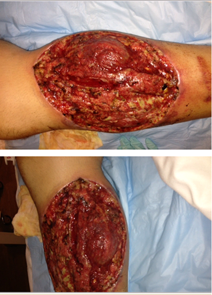 |
| Peripheral smear showing blasts and dysplastic neutrophils | 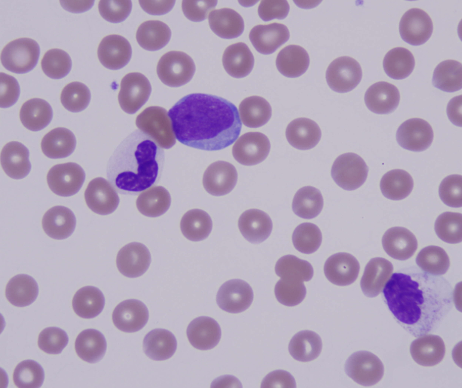 |
| BM biopsy showing increased immature cells and blasts in sheets | 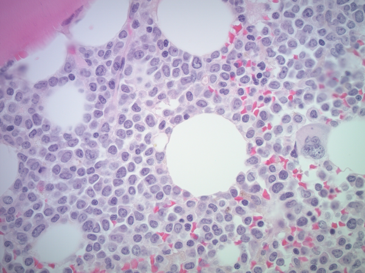 |
| Marrow aspirate showing atypical myeloid cells and numerous blasts | 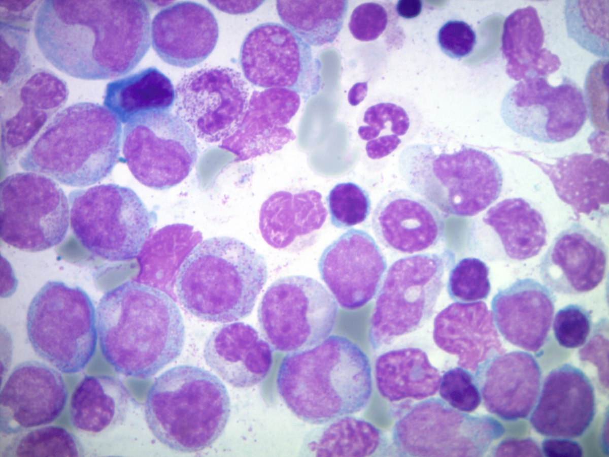 |
| Marrow aspirate showing atypical neutrophil and numerous maturing precursors | 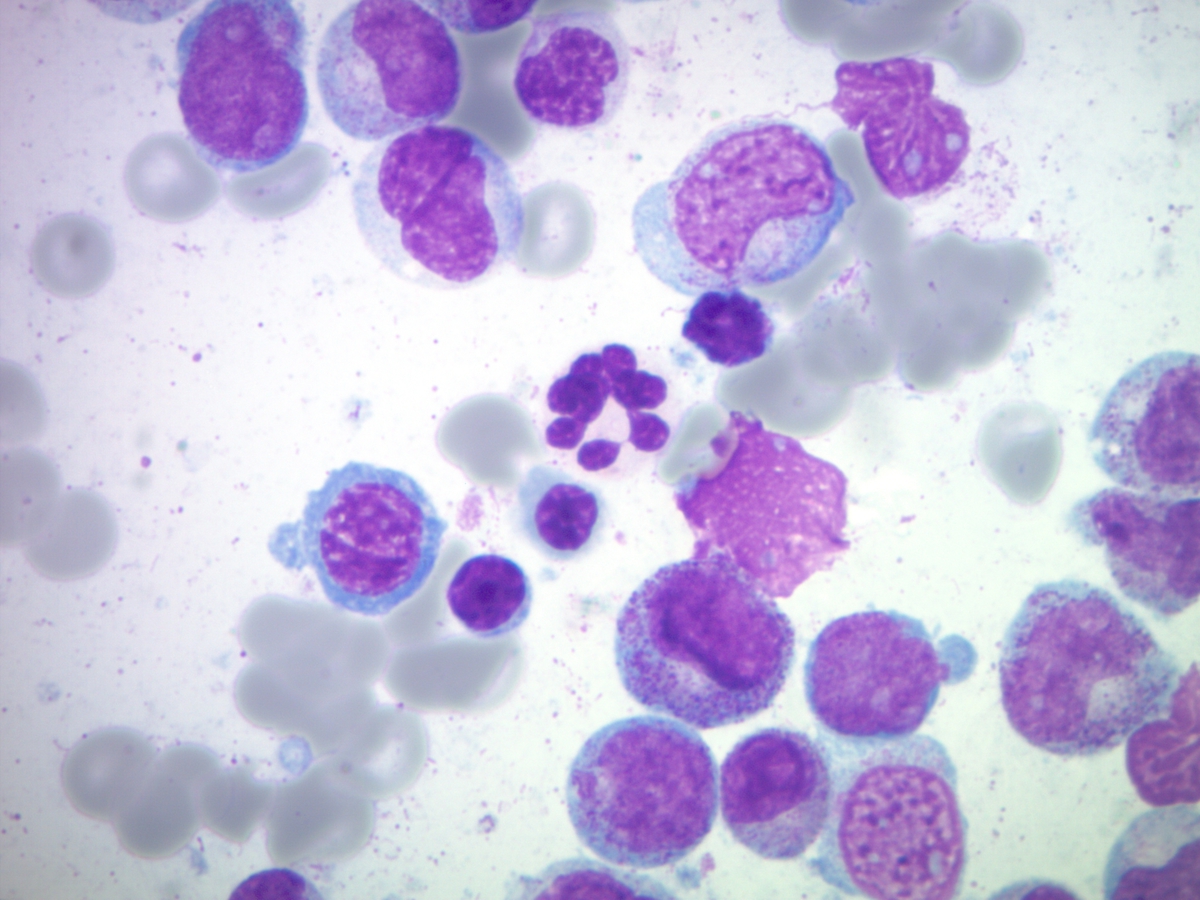 |