Institution: Brody School of Medicine at East Carolina University
Additional authors:Ridas Juskevicius, MD
Session: AML with myelodysplasia-related changes
HISTORY
The patient is an elderly female with history of pulmonary embolus on coumadin who presented with chronic normo-to-macrocytic anemia (Hgb 5.7-11.6g/L, MCV 99-117 fL) over a period of 4 years requiring multiple blood transfusions. Repeated B12 and folate studies were negative for deficiency and patient denied alcohol use. Patient was occasionally lymphopenic (0.9-2.0 k/uL) but neutrophil (1.8-5.2 k/uL) and platelet counts (185-370 k/uL) were within normal limits. Bone marrow biopsy was performed. Patient was subsequently placed on lenalidomide with reduced transfusion dependence.
DETAILS
Formalin fixed marrow biopsy section and aspirate from the right iliac spine demonstrated essentially normocellular marrow (30-40%) with trilineage hematopoiesis and slight erythroid hypoplasia. The majority of megakaryocytes contained hypo/mono-lobated nuclei. There was also hypolobation of some granulocyte precursors and some erythroid precursors had nuclear irregularities. Blasts were not increased.
IMMUNOHISTOCHEMISTRY AND FLOW CYTOMETRY
Flow cytometry performed on bone marrow aspirate demonstrated several populations of events corresponding to lymphocytes, monocytes and granulocytes. Only a minor population of events with dim CD45 expression and low side scatter characteristics corresponding to blasts was seen (less than 4%) with no phenotypically abnormal myeloid blasts identified.
CYTOGENETIC FINDINGS
46, XX, del(5)(q13q33)
MOLECULAR FINDINGS
Florescent in-situ hybridization with DNA probes (D5S23/D5S72, EGR1, D7S486, ETO and AML1) specific for the regions of chromosomes 5q15.2, 5q31, 7q31, 8q22, and 21q22 reveal a single copy of EGR1 (5q31) but no other abnormalities.
INTERESTING FEATURES
This case illustrated classic clinical, morphologic and cytogenetic features of MDS with isolated del(5q) or "5q- syndrome" - elderly female with isolated, severe macrocytic anemia with normocellular marrow containing slight erythroid hypoplasia, no increase in blasts and non- and hypolobated megakaryocytes.
First described in 1974 by van Den Berghe in 3 patients with refractory anemia and an interstitial deletion of the long arm of chromosome 5, the 5q- syndrome, is an independent subtype of MDS in the WHO classification system in which del(5q) is the sole cytogenetic abnormality. It is now established that 5q- syndrome belongs to collection of disorders known as "Ribosomopathies" in which genetic abnormalities cause impaired ribosome biogenesis and function, resulting in specific clinical phenotypes. Patients characteristically have a severe macrocytic anemia, normal/elevated platelets with hypolobulated megakaryocytes, and a relatively low rate of progression to acute myeloid leukemia compared with other types of MDS. Lenalidomide is highly effective for patients with the 5q- syndrome decreasing transfusion requirements in 76% of patients, and 61% of patients having a complete cytogenetic response.
RPS14 was identified as a 5q- syndrome gene in an RNA interference screen of each gene within the 5q- syndrome common deleted region. In patients with the 5q- syndrome, 1 allele of RPS14 is deleted, and haploinsufficient expression of RPS14 has been confirmed in patient samples. Decreased expression of RPS14 causes impaired erythropoiesis, with relative preservation of the other lineages. Heterozygous deletions of chromosome 5q in MDS are large, and haploinsufficiency for multiple genes probably contributes to the phenotype of the 5q- syndrome. However, in vitro and in vivo studies indicate that the erythroid defect, the aspect of the 5q- syndrome phenotype most analogous to DBA (Diamond-Blackfan anemia), is caused by RPS14 haploinsufficiency.
PROPOSED DIAGNOSIS
5q- syndrome
Myelodysplastic syndrome with isolated del(5q) (WHO 2008)
CONSENSUS DIAGNOSIS
Myelodysplastic syndrome with isolated del(5q)
| Bone marrow aspirate smear showing trilineage hematopoiesis with abnormal small and hypolobated megakaryocytes (x400, Wright-Giemsa) |  |
| Bone marrow biopsy section showing essentially normal cellularity with no abnormal infiltrates (x40, H&E) | 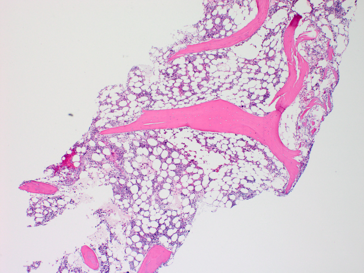 |
| Bone marrow biopsy section showing many abnormal small megakaryocytes with hypolobated nuclei (x400, H&E) | 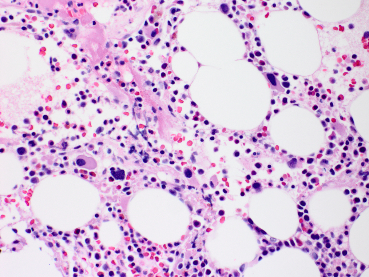 |
| Karyotype on bone marrow aspirate showing deletion of long arm of chromosome 5 (5q-)(arrowhead) | 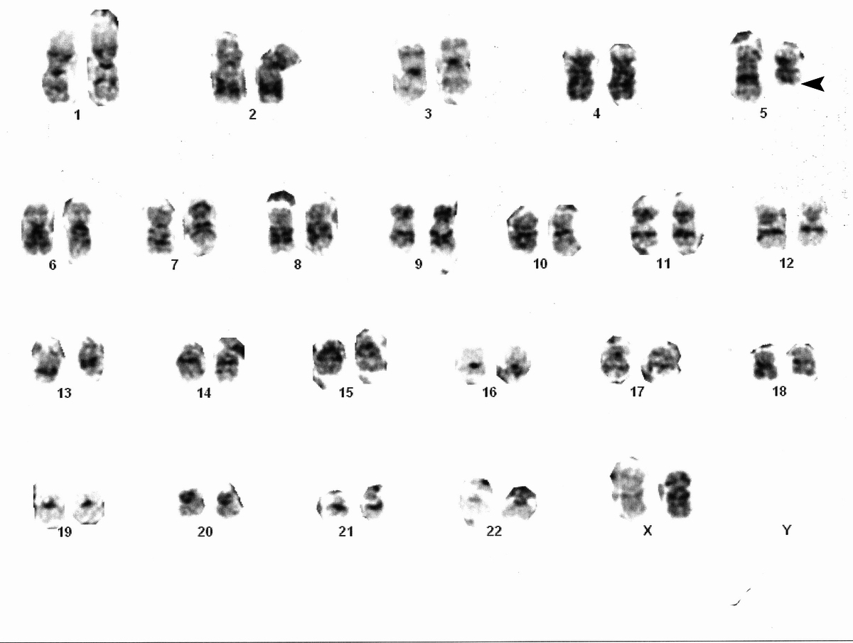 |
| FISH on bone marrow aspirate showing cells with 2 green and 1 red signals indicating deletion of long arm of chromosome 5 (VYSIS FISH probes to 5q31 EGR1 -red, 5p15.31 D5S7/D5S23 - green) |  |
| Selected bone marrow aspirate flow cytometry histograms showing no significant increase in blasts |  |