Institution: Department of Hematopathology, MD Anderson Cancer Center
Additional authors:Roberto N. Miranda, M.D., Keyur P. Patel, M.D, PhD, Jeffrey L.Jorgensen, M, Ph, Sa Wang, MD, Joseph D. Khoury, M, D Carlos Bueso Ramos, MD, PhD
Session: AML with myelodysplasia-related changes
HISTORY
81 year-old male, diagnosed with CMML-1 in July, 2010 with 80-90% bone marrow (BM) cellularity, trilineage dysplasia, 4% blasts and 13% monocytes. The CBC at diagnosis revealed Hgb 10.7 g/dl, plt 62 x 109/L and WBC 18.3 x 10⁹/L with 65% neutrophils, 8% metamyelocytes/myelocytes, 1% eosinophils, 11% lymphocytes, 15% of monocytes (2.4 K/UL) and 0% of blasts. The cytogenetic and molecular studies at diagnosis showed diploid karyotype, NRAS mutation at codon 13 and negative for JAK2 mutation. A CT scan showed splenomegaly.
His condition was stable for about 20 months and received supportive treatment and monthly monitoring. In April 2012, he was started on decitabine (20mg daily x 5 days) due to progressive leukocytosis (WBC up to 39.8 x 10⁹/L) with 1% blasts on peripheral blood (PB), 5% blasts in BM. He responded to the treatment initially, but developed leukopenia 2 months later and the dosage of decitabine was reduced. But he continued with pancytopenia and required RBC transfusions and received Neupogen, while Procrit was added later. The BM biopsy done in Sept. 2012 showed 10% blasts. Decitabine was discontinued due to significant pancytopenia. He was started on protocol “ARRAY-614,” a potent inhibitor of p38 mitogen-activated protein kinases and Tie2, on Nov. 2012.In Dec. 2012, the patient had leukocytosis and gradually PB showed 45% blasts. A BM biopsy was diagnosed with of “Acute Myeloid Leukemia (56% Blasts) transformed from Chronic Myelomonocytic Leukemia.” The diagnosis of “Systemic Mastocytosis with Associated Clonal Heamatological Non-mast Cell Lineage Disease” was also made. The patient and family decided on no further treatment and hospice care.DETAILS
Bone marrow biopsy site:
Left posterior iliac crestMethod of fixation: 10% buffered-formalinGross measurement: 1.1, 0.4, 0.2 cmMicroscopic description: Hypercellular marrow (95-100%) with decreased trilineage heamatopoietic cells; infiltrated by sheets of blasts with high nuclear/cytoplasmic ratio, scant basophilic cytoplasm. Multiple paratrabecular compact mast cell aggregates replacing 20% of marrow space were found.BM aspirate smear (December 2012) revealed 56% blasts, 7% atypical mast cells, 24% lymphocytes and 4% monocytes.The blasts were negative for myeloperoxidase and butyrate esterase cytochemical stains.IMMUNOHISTOCHEMISTRY AND FLOW CYTOMETRY
By immunohistochemistry, the mast cells reacted with mast cell tryptase and CD117.
By flow cytometry immunophenotype, the blasts expressed myeloid markers and were positive for CD13, CD15, CD33 (partial), CD34, CD38, CD45, CD49d, CD64 (partial), CD123, CD184, HLA-DR (partial) and TdT (partial). The blasts were negative for CD2, CD3 (surface and cytoplasmic), CD4, CD5, CD7, CD10, CD14, CD19, CD22, CD36, CD41, CD56, CD117 and myeloperoxidase.By flow cytometry immunophenotype, the mast cells showed an aberrant phenotype and expressed CD117, CD69, CD63, CD59, CD35 (partial) and CD25, and were negative for CD2, CD34.CYTOGENETIC FINDINGS
7/7/2010: Diploid male karyotype (46,XY[20]); negative for BCR/ABL1 gene arrangement by FISH.
12/8/2010: diploid male karyotype (46,XY[20]).3/14/2012: diploid male karyotype (46,XY[20]).7/12/2012: diploid male karyotype (46,XY[7]).9/7/2012: diploid male karyotype (46,XY[20]).10/24/2012: diploid male karyotype (46,XY[20]).12/19/2010: no mitotic cells recovered.MOLECULAR FINDINGS
7/7/2010:
Positive for NRAS mutation (codon 13, G13V). Negative BCR-ABL fusion transcript by real-time PCR; negative for JAK2 mutation at codon 617; negative for FLT3 (ITD and codon 835/836 mutation), KIT, NPM1 and CEBPA mutations. 12/8/2010: Positive for NRAS mutation (codon 13, G13V). Negative for FLT3 mutation.3/14/2012: Positive for NRAS mutation (codon 13, G13V). Negative for IDH1, FLT3, KRAS, IDH2HRM.7/12/2012: Negative for KRAS, NRAS mutations.9/7/2012: Negative for KRAS, NRAS and JAK2 mutations.10/10/2012: Negative for KRAS, NRAS mutations.10/24/2012: Negative for KRAS, NRAS mutations.12/5/2012: Positive for NRAS mutation (codon 13, G13V). Negative for KRAS mutation.12/19/2012: Positive for NRAS mutation (codon 13, G13V); negative for KRAS mutation. The molecular study for KIT mutation (D816V) is in progress.INTERESTING FEATURES
CMML is a myelodysplastic/myeloproliferative neoplasm. RAS mutation has been associated with CMML. We have shown that either KRAS or NRAS is a poor prognostic marker for CMML patients. Although this patient has diploid karyotype and the disease was stable for about 20 months, he developed AML and the outcome was poor.
Interestingly, the patient was tested positive for NRAS at the original diagnosis of CMML, and continued to be positive for NRAS before the initiation of the decitabine treatment. However, he became NRAS negative after started the decitabine treatment, and remained negative even when the BM blasts counts increased from 4% to 10%. Only at the time of development of AML, the NRAS became positive again. Therefore, there might be a possible association between the decitabine treatment and the inhibition the NRAS mutated clone, and when such inhibitory effect is lost, the progression to AML could occur.Another interesting feature of this case was the development of systemic mastocytosis when the patient progressed to AML. The hematological disorder most commonly associated with systemic mastocytosis is CMML. Our patient did not have systemic mastocytosis during the CMML phase but systemic mastocytosis was identified when he developed AML.PROPOSED DIAGNOSIS
Acute Myeloid Leukemia (56% Blasts) Transformed from Chronic Myelomonocytic Leukemia.
Systemic Mastocytosis with Associated Clonal Hematologic Non-mast Cell Lineage Disease.CONSENSUS DIAGNOSIS
Acute myeloid leukemia with myelodysplasia-related changes, arising from chronic myelomonocytic leukemia, with systemic mastocytosis with associated clonal hematologic non-mast cell lineage disease
| Figure 1 | 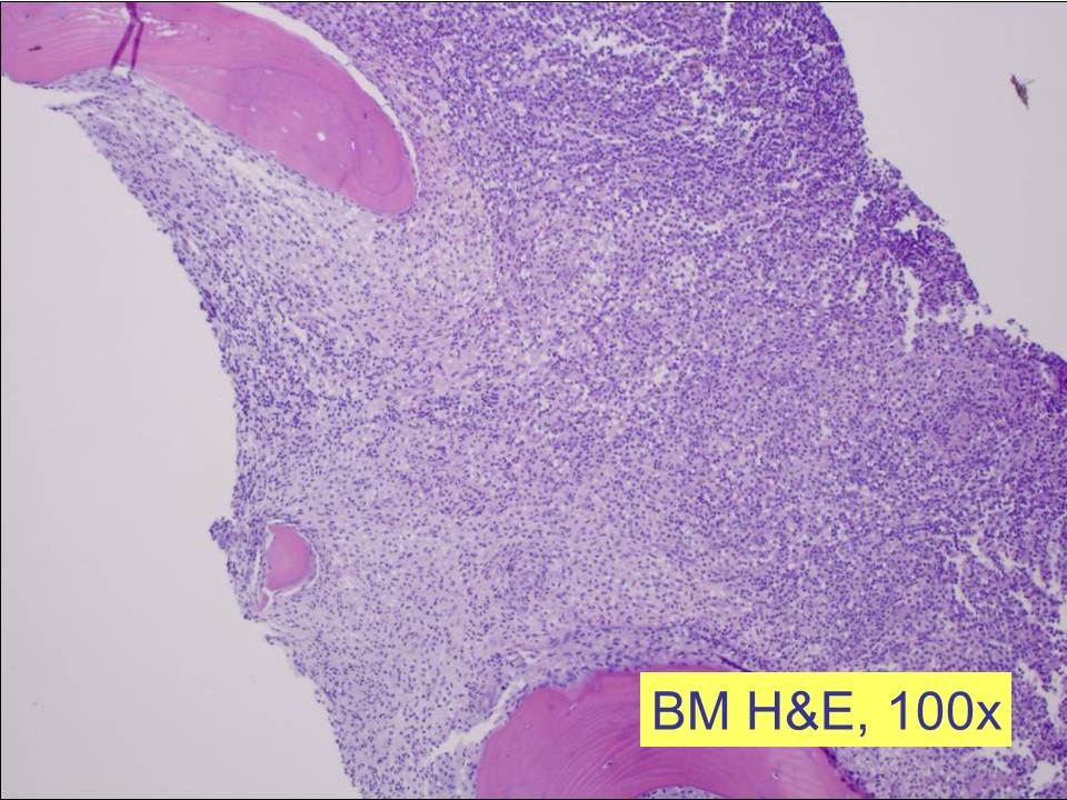 |
| Figure 2 | 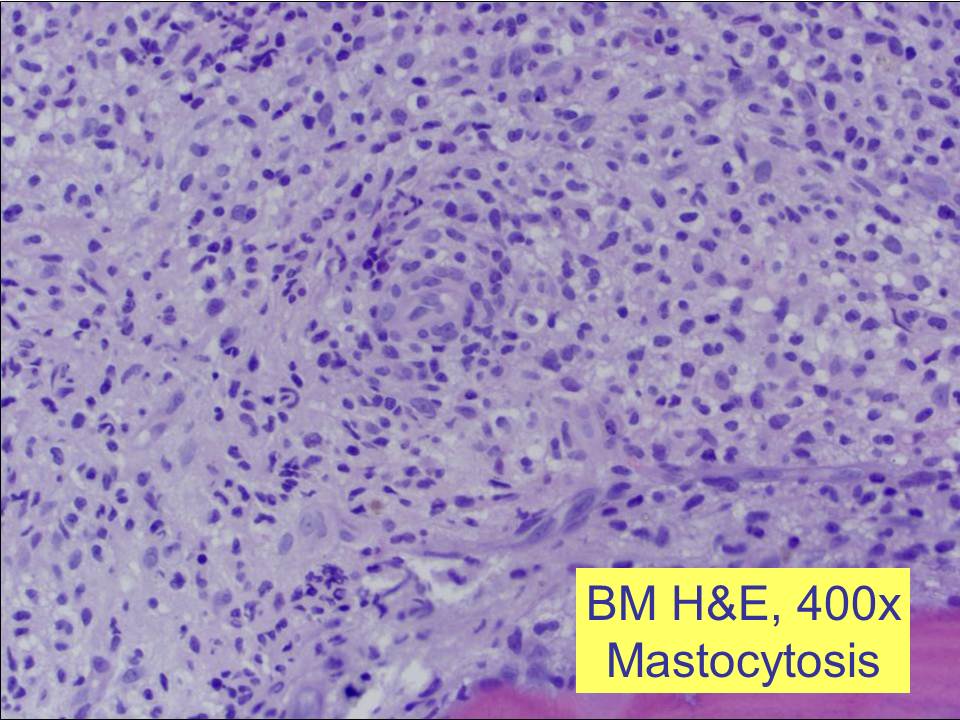 |
| Figure 3 |  |
| Figure 4 | 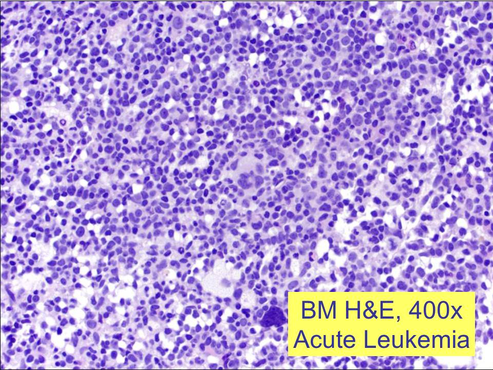 |
| Figure 5 |  |
| Figure 6 | 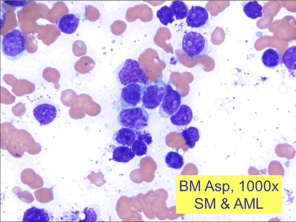 |
| Figure 7 |  |
| Figure 8 |  |
| Figure 9 | 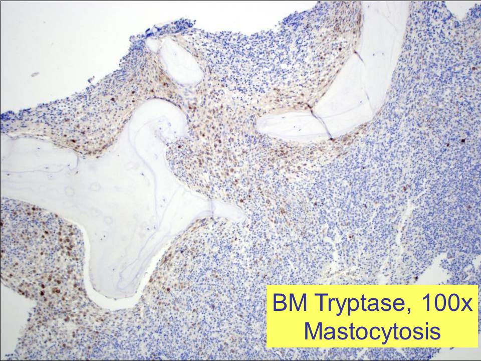 |
| Figure 10 | 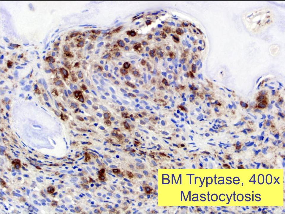 |
| Figure 11 | 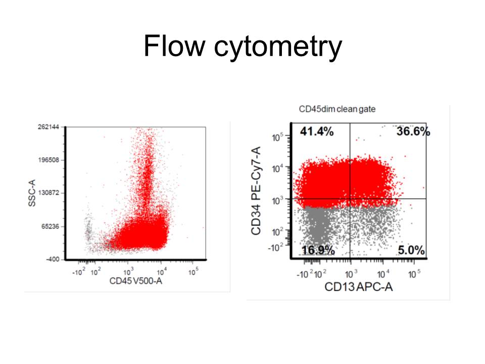 |
| Figure 12 | 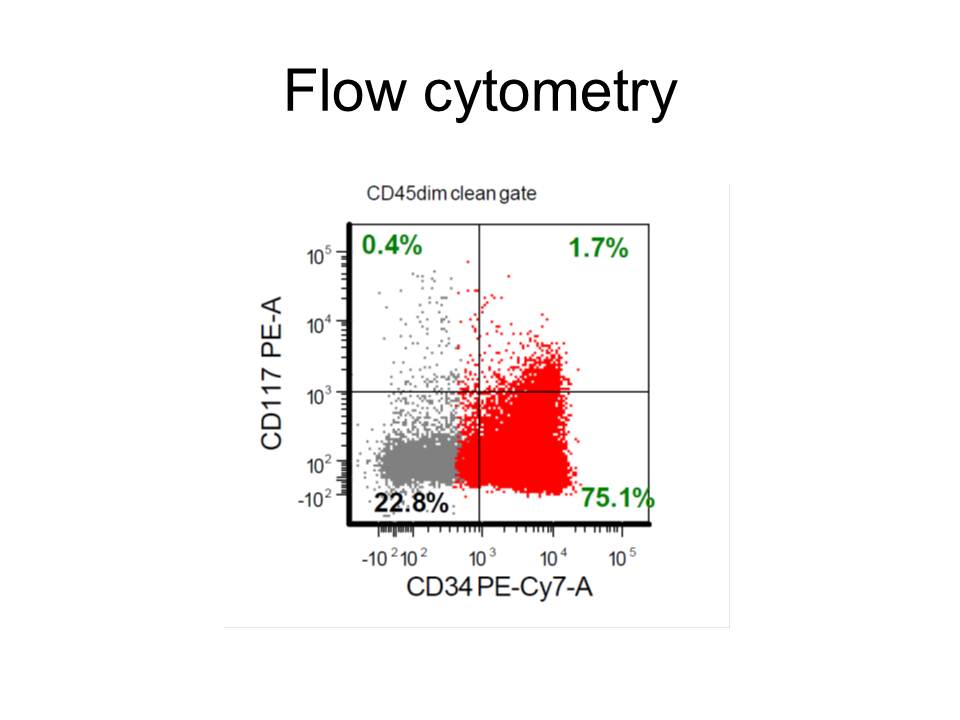 |
| Figure 13 | 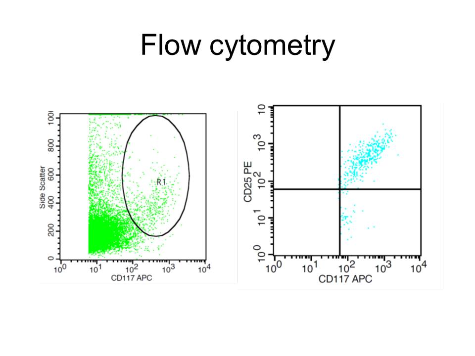 |