Institution: MD Anderson Cancer Center
Additional authors:Yang O. Huh, MD
Session: Erythroleukemia and megakaryoblastic AML and mimics
HISTORY
68-year-old male with a history of mantle cell lymphoma, status post allogeneic stem cell transplant on 3/31/2011, developed pancytopenia accompanied by headache. The patient was not managed with erythropoietin or other growth factors Chief complaint: fatigue Physical examination: Unremarkable Laboratory findings - H/H: 9.5/27.7, WBC: 1.4, platelet: 18, absolute neutrophilic count: 0.44
DETAILS
Site: Left posterior iliac crest Method of fixation: formalin-fixed Microscopic pathology - Biopsy: Hypercellular bone marrow (80%) with increased immature cells with large vesicular nuclei with prominent nucleoli, interstitial and focally forming sheets involving approximately 80% of marrow cellularity - Touch preparation: Markedly increased medium to large size abnormal proerythroblasts (40%) with fine chromatin, one to several large nucleoli, and abundant basophilic cytoplasm containing fine vacuoles - PAS stain: Positive, finely granular and block positive in blasts - Smear: poor quality - Myeloperoxidase: negative
IMMUNOHISTOCHEMISTRY AND FLOW CYTOMETRY
Positive stains - Glycophorin A (30-50%), E-cadherin, and CD11c (dim, in subset) Negative stains - CD3, CD10, CD30, CD43, CD61, CD68 (PG-M1), ALK1, BCL-2, BCL-6, cyclin D1, factor VIII, keratin, lysozyme, PAX-5 Flow Cytometry - Non-contributory due to poor quality of aspirate specimen
CYTOGENETIC FINDINGS
58~59,XY,-3,-7,-9,-10,-13,-13,-16,-17,-19,-20,+21,-22,+2mar[cp3]/ 46,XY,t(2;15)(p23;q26)[1]/46,XY[26] FISH - IGH@/CCND1: negative - Probe for chromosome 5: Aneusomy 5 - Probe for chromosome 7: Aneusomy 7
MOLECULAR FINDINGS
JAK2 codon 617: wild type IDH1/IDH2: wild type NPM1 exon12: wild type FLT3: wild type BCR-ABL1: negative
INTERESTING FEATURES
Therapy-related acute myeloid leukemia (proerythroblastic) is rare. Cases of pure erythroid leukemia express 1 or more erythroid antigens (Glycophorin A, hemoglobin A or E-cadherin). In addition, complex karyotype was found, commonly seen in therapy-related setting. The differential diagnosis of the pure erythroid leukemia includes other types of AML, acute lymphoblastic leukemia, lymphoma, plasma cell myeloma among others.
PROPOSED DIAGNOSIS
- Therapy-related acute myeloid leukemia with prominent erythroblastic differentiation
- No evidence of mantle cell lymphoma
CONSENSUS GROUP: ADDITIONAL INFORMATION/STUDIES
The immature erythroblasts were E-cadherin positive; glycophorin positive in 30-50% cells. Total immature erythroblasts were 60%. The patient was treated with low-dose cytarabine and clofarabine, achieved a very short partial response. Treatment was changed to Vidaza and tosedostat, but patient had no response and died. Total survival: 5 months.
CONSENSUS DIAGNOSIS
Therapy-related myeloid neoplasm; consistent with acute erythroid leukemia (pure erythroid leukemia)
| Bone marrow biopsy 4x | 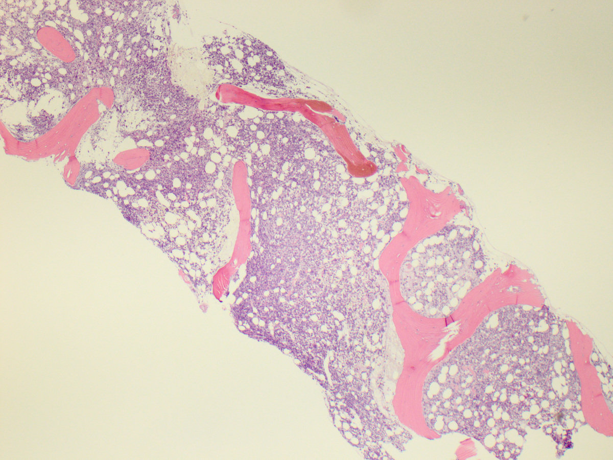 |
| Bone marrow biopsy 40x | 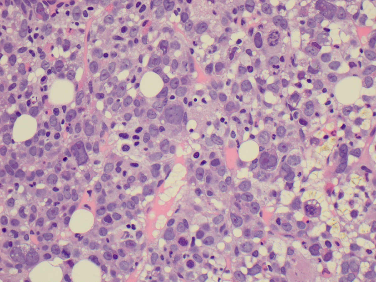 |
| Bone marrow touch preparation 100x | 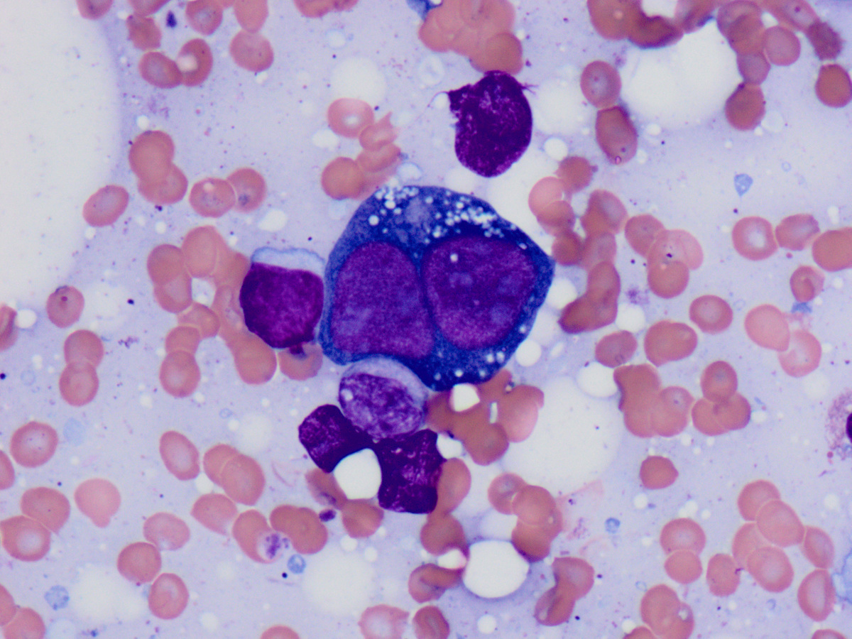 |
| Bone marrow touch preparation, PAS stain, 100x |  |
| Bone marrow biopsy, glycophorin A stain, 40x |  |
| Bone marrow biopsy, E-cadherin stain |  |