Institution: Kingston General Hospital, Queen's University
Additional authors:Michael Rauh, MD, FRCPC
Session: AML with recurrent genetic abnormalities Part I
HISTORY
15 year old male with history of spina bifida. He presented to hospital with normocytic anemia and marked thrombocytopenia with 72% circulating blasts. The morphological appearance and immunophenotype were that of myeloid blasts. He was began treatment with the COG protocol AAML1031. On day 28, a repeat bone marrow was performed and revealed less than 5% blasts but had a significant increase in mast cells and plasma cells. The patient also had clinical symptoms of systemic mastocytosis including urticaria and dermatographism.
DETAILS
Initial bone marrow:
Bone marrow aspirate and biopsy, posterior iliac crest, side not specified. Biopsy fixed in AZF, decalcification in RDO. Aspirate reveals 65% intermediate to large blasts with irregular nuclei, fine chromatin, occasional small nucleoli and rare thin Auer rods. Some hypogranular and hyposegmented neutrophils are present. There is also a mild increase in eosinophil precursors. The biopsy is tangential and consists almost exclusively of periosteal tissue and cortical bone with only a tiny focus of marrow showing blasts and a few eosinophils.
Day 28 bone marrow:
Bone marrow aspirate only. The aspirate contains only 2% blasts by morphology. There is an increase in mast cells comprising up to 25% of the cellularity with loose aggregates of mast cells present in the particles. There is also an increase in mature appearing plasma cells. Mildly reduced erythropoiesis and granulopoiesis.
IMMUNOHISTOCHEMISTRY AND FLOW CYTOMETRY
Initial bone marrow:
Flow cytometry reveals 59% blasts expressing CD34, CD117, CD13, HLA-DR and myeloperoxidase with partial expression of CD19. Biopsy insufficient for IHC assessment.
Day 28 bone marrow:
Flow cytometry reveals less than 1% CD34 positive blasts. No biopsy obtained for IHC studies.
CYTOGENETIC FINDINGS
Initial bone marrow:
46,XY, t(8;21)(q22;q22) detected in 10 of 10 metaphases.
This was confirmed by FISH in 191 of 230 interphase nuclei.
Day 28 bone marrow:
46,XY in 18 metaphases.
FISH analysis revealed t(8;21) in 60/232 (26%) of interphase nuclei.
MOLECULAR FINDINGS
Initial bone marrow:
Not performed.
Day 28 bone marrow:
A Thr411fs mutation in the KIT gene was detected by RT-PCR and sequencing of exon 8 of the KIT gene.
INTERESTING FEATURES
Although KIT mutations may be present in up to a quarter of cases of t(8;21)(q22;q22) AML, the concurrent or subsequent development of systemic mastocytosis has only rarely been reported. There is a suggestion that the leukemic blasts and mast cells may derive from a common malignant progenitor. This is also suggested in our case with 26% of nuclei having the t(8;21) translocation in the day 28 sample, similar to the percentage of mast cells and there being only 2% blasts in this aspirate sample.
PROPOSED DIAGNOSIS
Acute myeloid leukemia with t(8;21)(q22;q22); RUNX1-RUNX1T1 with development of systemic mastocytosis post induction chemotherapy.
CONSENSUS DIAGNOSIS
Acute myeloid leukemia with t(8;21)(q22;q22); RUNX1-RUNX1T1, and development/detection of systemic mastocytosis following therapy
| Initial peripheral blood blasts |  |
| Initial peripheral blood blasts 60x | 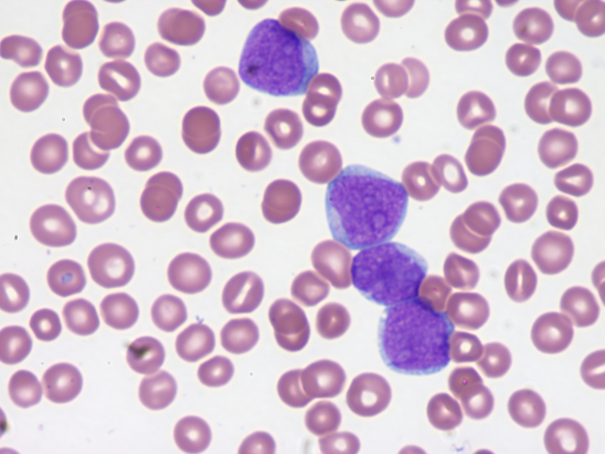 |
| Initial bone marrow aspirate 10x | 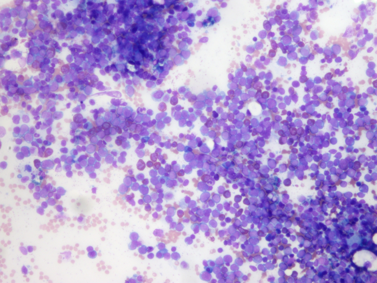 |
| Initial bone marrow aspirate 20x | 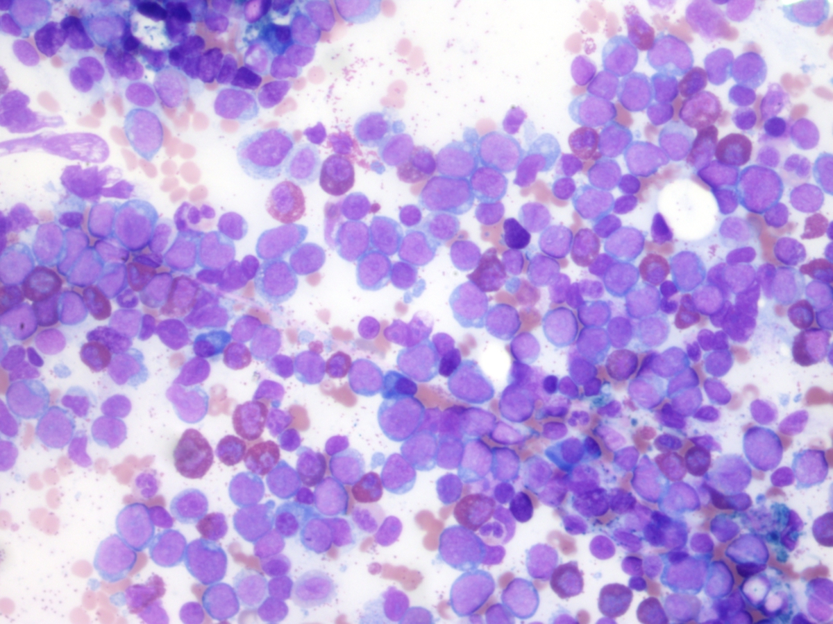 |
| Initial bone marrow aspirate 60x |  |
| Initial bone marrow biopsy 2x |  |
| Initial bone marrow biopsy 40x | 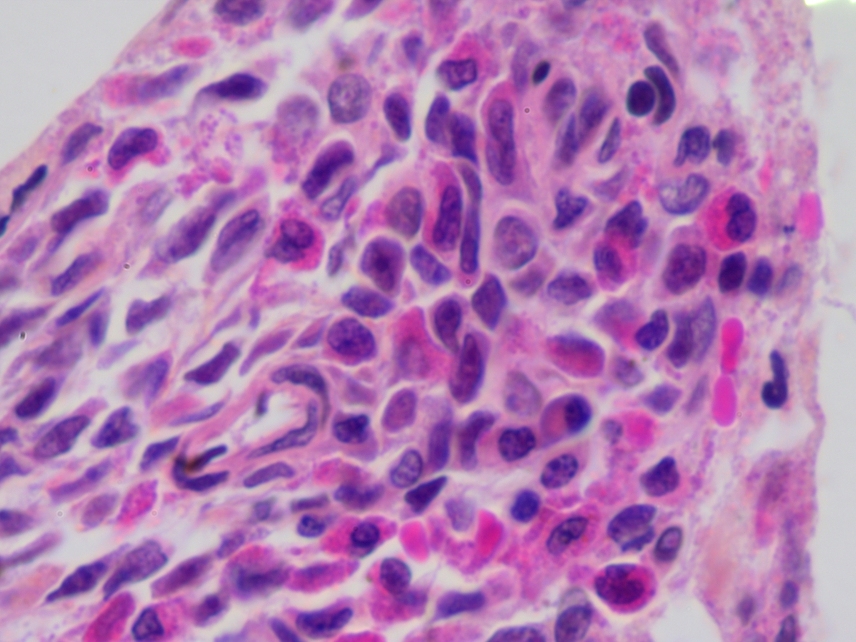 |
| Day 28 bone marrow aspirate showing mast cells | 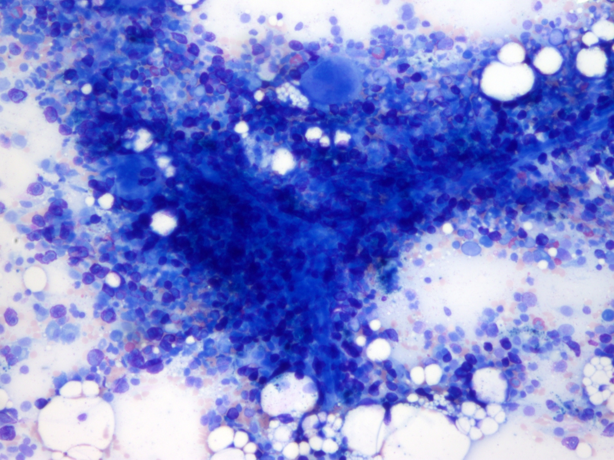 |
| Day 28 bone marrow aspirate showing mast cells |  |
| Day 28 bone marrow aspirate showing mast cells |  |