Institution: Brody School of Medicine at East Carolina University
Additional authors:Jonathan D. Boyd, MD; Jun Zhang, MD; Ronald Mageau, MD
Session: Myeloid and lymphoid neoplasms with eosinophilia and abnormalities of PDGFRA, PDGFRB, or FGFR1
HISTORY
Caucasian male in his early 50s presented with severe back pain, night sweats, weight loss and bilateral lower extremity weakness for two weeks. Physical examination showed splenomegaly. The complete blood count (CBC) initially showed normal white cell count (4,000/µl) with mild eosinophilia (900/µl). Computed tomography (CT) showed bilateral pelvic lymphadenopathy and splenomegaly. 2-D echocardiogram was normal. Lymph node aspiration was performed followed by bone marrow biopsy.
After diagnosis, the patient was treated with conventional induction chemotherapy with daunorubicin and cytarabine. Day 14 BM was acellular with focal fibrosis but no evidence of AML. Day 30 BM was hypercellular with eosinophilia and fibrosis but no increase in blasts, consistent with chronic myeloproliferative neoplasm. Subsequently, the patient developed worsening of the back pain and lower extremity numbness. CBC at this time showed leukocytosis (9,900/µl) with absolute eosinophils of 3,000/µl. Magnetic resonance imaging (MRI) of the spine showed epidural/ paraspinal tumor extending from T5 - L5 vertebrae with moderate cord compression at T11. A T11 laminectomy with epidural mass biopsy showed a myeloproliferative neoplasm with eosinophilia and fibrosis similar to BM. Imatinib was started at 400 mg/day. In two days the absolute eosinophils decreased to 300/µl. Neurological symptoms resolved in one week. Repeat MRI in two months showed near complete regression of the epidural tumor. The patient has been on imatinib for 10 months, and remains in complete remission.DETAILS
Formalin fixed paraffin-embedded H&E stained marrow biopsy sections from the posterior superior illiac spine demonstrated markedly hypercellular marrow (95%) with distinct areas containing extensive diffuse infiltrate of blasts (>80%). The blasts were intermediate to large in size and have round to oval slightly irregular nuclei with prominent nucleoli. There was significant associated eosinophilia. No normal hematopoietic elements were seen in the areas of the marrow containing the blast infiltrate. Other morphologically distinct areas seen on this biopsy had only rare blasts but showed extensive eosinophilia with other more mature myeloid elements and occasional megakaryocytes seen. The few megakaryocytes that were seen had unremarkable morphology. Reticulin stain showed marked diffuse increase in reticulin fibrosis.
Touch preparations showed predominantly peripheral blood with only rare bone marrow cells, including few blasts. Cytochemical staining with butyrate esterase and myeloperoxidase (MPO) was performed with functional controls and showed rare cells to be positive for MPO and no cells staining for butyrate esterase.Peripheral blood showed abnormal eosinophils with atypical nuclei and features of degranulation and rare circulating blasts.IMMUNOHISTOCHEMISTRY AND FLOW CYTOMETRY
The flow cytometric immunophenotyping on the lymph node aspirate revealed a significant population of events with dim CD45 staining and low side scatter, corresponding to blasts. The blasts had an immature immunophenotype (CD11b dim, CD33, CD34, CD38, dim HLA-DR) most consistent with myeloid origin. There was dim expression of CD5, but no coexpression of TdT, cytoplasmic CD3 or any other B or T-cell antigens by the blasts.
Immunohistochemical stains on the bone marrow biopsy sections demonstrated that blasts were CD34+/ CD117+/ CD43+ and were negative for Lysozyme and CD68.CYTOGENETIC FINDINGS
Conventional cytogenetic study was attempted but was not successful
MOLECULAR FINDINGS
Florescent in situ hybridization (FISH) using VYSIS 4q12 tricolor rearrangement probe revealed findings consistent with FIP1L1-PDGFRα fusion (1G1A signal indicating loss of LNX [red] signal and one normal fusion signal containing intact LNX [red] probe). In addition, FISH was negative for inv(16), BCR-ABL fusion and abnormalities at 5q33 and 8p11.
INTERESTING FEATURES
Myeloproliferative neoplasms (MPN) with PDGFRα rearrangements usually present as chronic eosinophilic leukemia with end organ damage, but rarely may present as AML. Presentation of MPN with FIP1L1-PDGFRα fusion as an extramedullary AML (myeloid sarcoma) is exceedingly rare. To the best of our knowledge, this is the first case of this entity presenting as extramedullary AML in a lymph node, clinically mimicking lymphoma. This case is also very interesting morphologically showing two clearly defined areas in the bone marrow biopsy containing diffuse infiltrate of blasts and adjacent area containing essentially no blasts but showing features of chronic myeloproliferative neoplasm with fibrosis and eosinophilia. After AML induction therapy day 30 bone marrow sample showed no blast infiltrates but recurrent chronic myeloproliferative neoplasm with eosinophilia and fibrosis. Similar morphologic features were present in the latter laminectomy specimen of the paraspinal mass.
FIP1L1-PDGFRα fusion protein is a constitutively active tyrosine kinase, which is extremely sensitive to the tyrosine kinase inhibitor imatinib (100-fold more sensitive than BCR-ABL). Given this therapeutic implication and the fact that these tumors can present as AML in extramedullary sites, as illustrated by our case, the importance of screening for PDGFRα rearrangement in patients presenting with AML with eosinophilia and/ or extramedullary involvement cannot be overemphasized.PROPOSED DIAGNOSIS
Blast transformation (acute phase) of a chronic myeloproliferative neoplasm with PDGFRα rearrangement (FIP1L1-PDGFRα fusion)
CONSENSUS DIAGNOSIS
Myeloid neoplasm with PDGFRA rearrangement (FIP1L1-PDGFRA fusion), presenting with acute myeloid leukemia, possibly evolving from myeloproliferative neoplasm
| FNA of the pelvic lymph node showing population of blasts in a background of small mature lymphocytes. More mature myeloid/ eosinophilic precursor is seen in lower right (x100, Diff-Quik stain) | 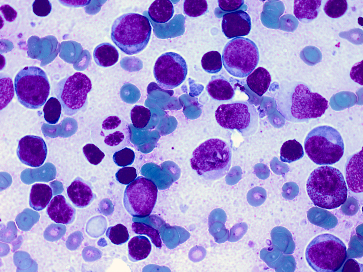 |
| Flow cytometry of the FNA from pelvic lymph node showing population of CD33+ blasts with no expression of CD3 or CD19 | 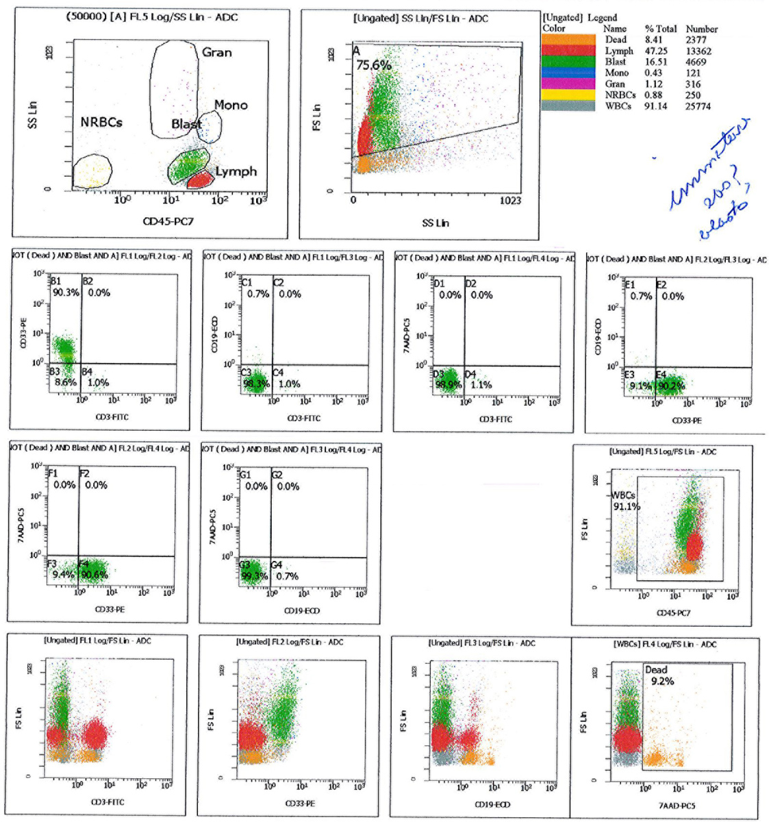 |
| Flow cytometry of the FNA from pelvic lymph node showing that blasts have no expression of lymphoid markers except dim CD5 | 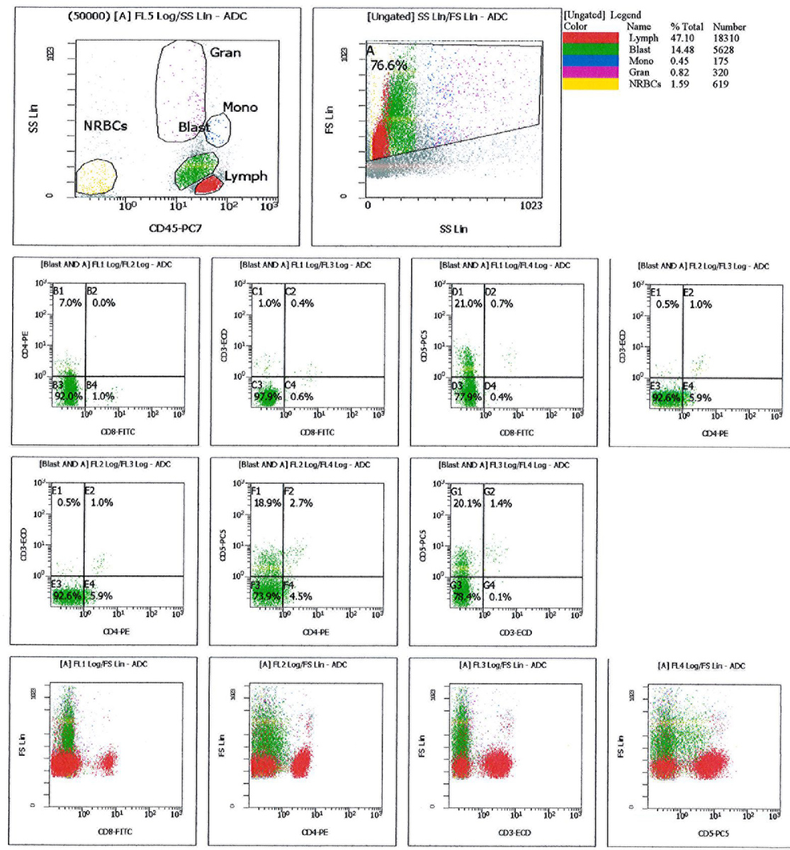 |
| Flow cytometry of the FNA from pelvic lymph node showing that blasts have dim expression of CD11b but no expression of CD56 or CD16 | 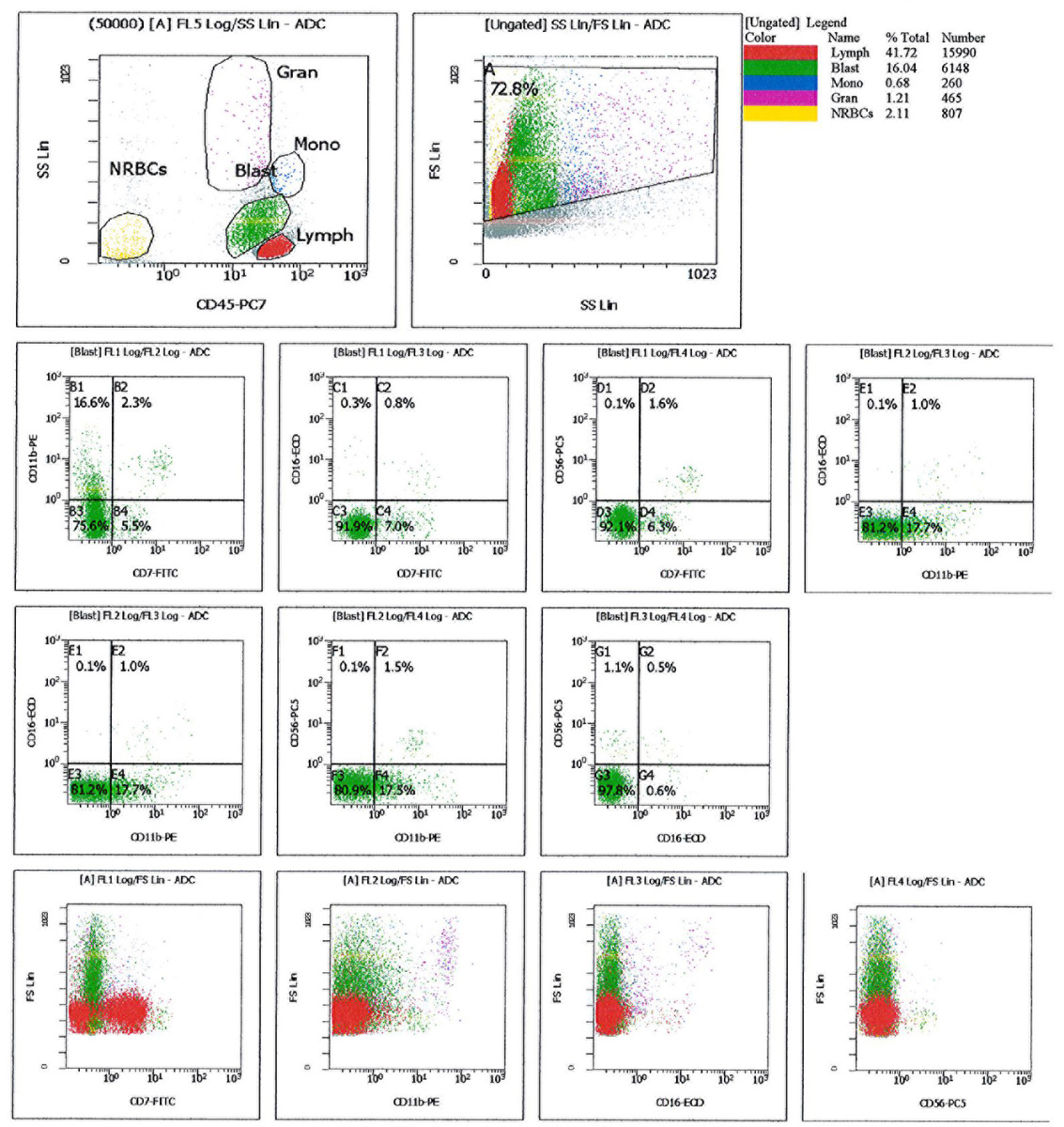 |
| Flow cytometry of the FNA from pelvic lymph node showing that blasts are CD34+ and have borderline expression of HLA-DR | 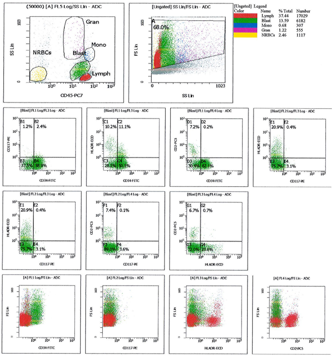 |
| Flow cytometry of the FNA from pelvic lymph node showing that blasts have no expression of other myelomonocytic markers |  |
| Peripheral blood smear with some eosinophils showing abnormal nuclear features. Rare blast cells were also seen (x400, Wright-Giemsa) |  |
| Bone marrow biopsy sections showing markedly hypercellular marrow with infiltrate of immature cells on the left and more eosinophilic fibrotic area on the right (x40, H&E) |  |
| BM biopsy sections showing interface between sheets of blasts on the left and fibrotic area with more mature cells and eosinophils on the right (x40, H&E) |  |
| BM biopsy sections showing area composed of diffuse extensive infiltrate of blasts and eosinophils (x400, H&E) | 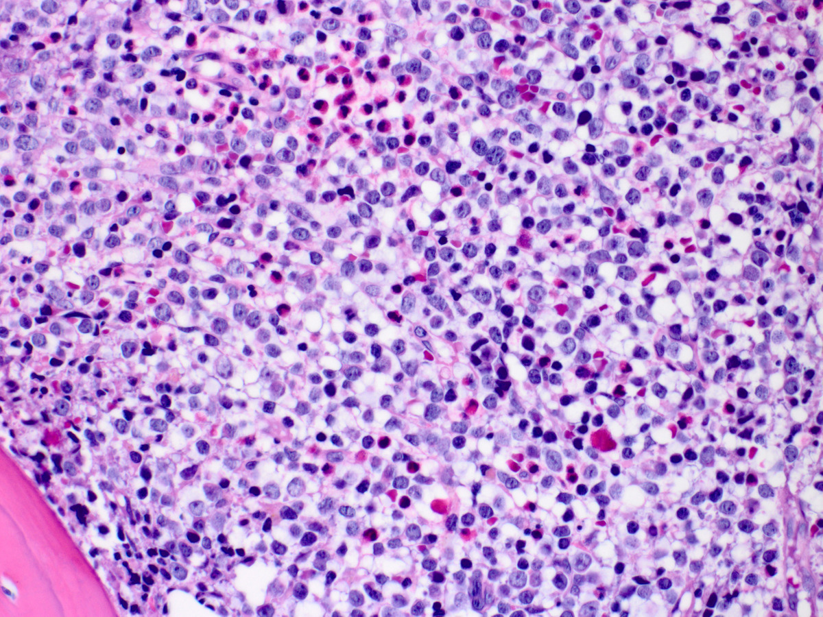 |
| BM biopsy sections showing area composed of fibrotic stroma, more mature myeloid cells including eosinophils (x400, H&E) |  |
| Reticulin stained bone marrow biopsy section showing extensive fibrosis (x40) |  |
| CD34 stain on bone marrow biopsy showing interface between area of blast transformation (left) and chronic phase (right) (x40) |  |
| CD117 stain on the BM biopsy showing positivity in most blast cells (x200) |  |
| FISH using Vysis LSI 4q12 tricolor rearrangement probe on peripheral blood sample showing loss of red signal (LNX) indicating fusion between FIP1L1 and PDGFRA |  |
| Magnetic resonance imaging (MRI) of the spine showed epidural/ paraspinal tumor extending from T5 - L5 vertebrae with moderate cord compression at T11 (A, red arrows indicate tumor infiltrate). Repeat MRI two months after starting Imatinib therapy showed near complete regression of the epidural tumor (B). |  |