Institution: Pathology Associate of Albuquerque
Additional authors:Alexis Bakhirev, Sever Cordelia
Session: AML with recurrent genetic mutations Part II
HISTORY
This is a 49-year-old male with no significant past medical history who came in with pancytopenia and fatigue for the last few weeks. He also had some fevers, chills, upper respiratory symptoms, headaches, new oral lesions and cough. A bone marrow was performed.
DETAILS
Bone marrow biopsy site: Right posterior iliac crest
Fixation: Standard bone marrow fixation by AZF
Gross: 1.3 cm trephine biopsy and clot sections
Microscopic pathology:
Peripheral blood: Pancytopenia with circulating blasts and dysplastic neutrophils
WBC: 2.2 x 10^3/mm3
Neut: 26 %
RBC: 2.31 x 10^6/mm3
Lymph: 43 %
Hgb: 9 gm/dL
Var Lymph: 14 %
HCT: 26 %
Mono: 6 %
Blast: 11 %
MCV: 110 fL
MCHC: 34.9 gm/dL
RDW: 17.8 %
Plt: 52 x10^3/mm3
Bone marrow aspirate smears contain 40% blasts without morphological features of myeloid differentiation. Background granulocytes show dysplastic features.
On the trephine biopsy and clot sections, there is atypical interstitial infiltrate composed of small to intermediate, slightly irregular cells with immature chromatin. Residual hematopoiesis is markedly decreased.
IMMUNOHISTOCHEMISTRY AND FLOW CYTOMETRY
This is an unusual immunophenotype which is difficult to classify. Flow cytometric analysis identifies an increased number of blasts (46%) in this bone marrow aspirate specimen that express dim CD45, CD34, and CD117 (major subset). Lineage classification was complicated by the either non-specific or possible expression of CD4, CD7, CD10, and CD13 in a small subset of blasts. Other myeloid associated, monocytic, megakaryocytic, and erythroid lineage antigens are indeterminate or negative. Lineage specific lymphoid markers are negative. A myeloperoxidase cytochemical stain is also negative. By immunohistochemistry, blast infiltrate is positive for CD34; CD123 and TCL1a are largely negative in blasts, which helps to rule out blastic plasmacytoid dendritic cell neoplasm.
CYTOGENETIC FINDINGS
Cytogenetic analysis: 46,XY[20]
FISH analysis:
There is no evidence for the RUNX1T1/RUNX1 fusion associated with the translocation t(8;21)(q22;q22) in the cells examined, using the RUNX1T1/RUNX1 dual color, dual fusion probe system.
There is no evidence for the PML/RARA fusion associated with the translocation t(15;17) in the cells examined, using the LSI PML/RARA dual color, dual fusion probe system.
There is no evidence for rearrangement of the CBFB gene, which may be associated with the inversion 16(p13q22), in the cells examined, using the dual color, break apart probe system.
MOLECULAR FINDINGS
Negative for the FLT3 Internal Tandem Duplication
Negative for the FLT3 D835 Variant
Negative for a NPM1 exon 12 mutation
INTERESTING FEATURES
By flow cytometry, immunohistochemistry, and special stains blasts do not show a definite lineage specific marker. However, presence of CD117 and possible subset CD13, both typically seen with myeloid leukemias albeit not lineage specific, along with morphologic findings of myeloid dyspoiesis suggest myeloid origin. This case brings up a few questions:
1. MPO is considered to be the only specific myeloid marker by WHO classification. In routine practice, if blasts are positive for a variety of myloid marker (including monocytic markers) with appropriate morphology, it would be classified as acute myeloid leukemia. Is MPO the only absolute crierior for myeloid differentiation?
2. This case is probably best classifed as acute undifferentiated leukemia. Though it also fits the description of acute myeloid leukemia with minimal differentition which the diagnostic criteria provided by WHO is somewhat conflicting and confusing (Page 130, WHO 2008)
3. Could background dyspoiesis be used as one of criterior to suggest myeloid origin? Is comment of "suggestive of myeloid origin" treatment-relevant?
PROPOSED DIAGNOSIS
Acute undifferentiated leukemia
CONSENSUS GROUP: ADDITIONAL INFORMATION/STUDIES
Immunostains performed by the consensus group show positive staining for MPO, lysozyme, and CD11c in a subset of cells.
These findings, along with background megakaryocytic dysplasia, support myeloid differentiation (AML with MRC).
CONSENSUS DIAGNOSIS
Acute myeloid leukemia with myelodysplasia-related changes
| Flow Cytometry 1 | 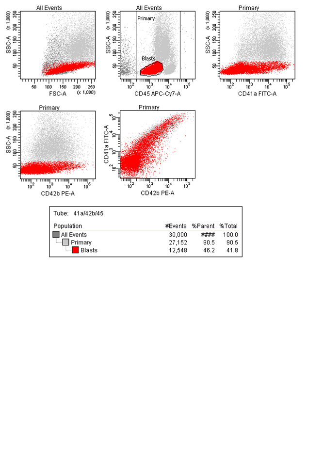 |
| Flow Cytometry 2 |  |
| Flow Cytometry 3 |  |
| Flow Cytometry 4 | 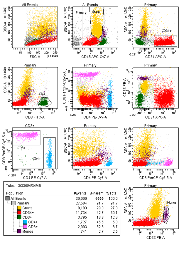 |
| Flow Cytometry 5 | 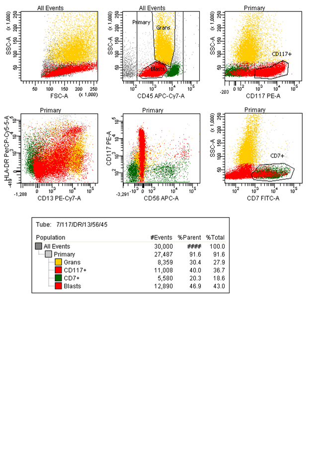 |
| Peripheral blood: dysplastic neutrophils | 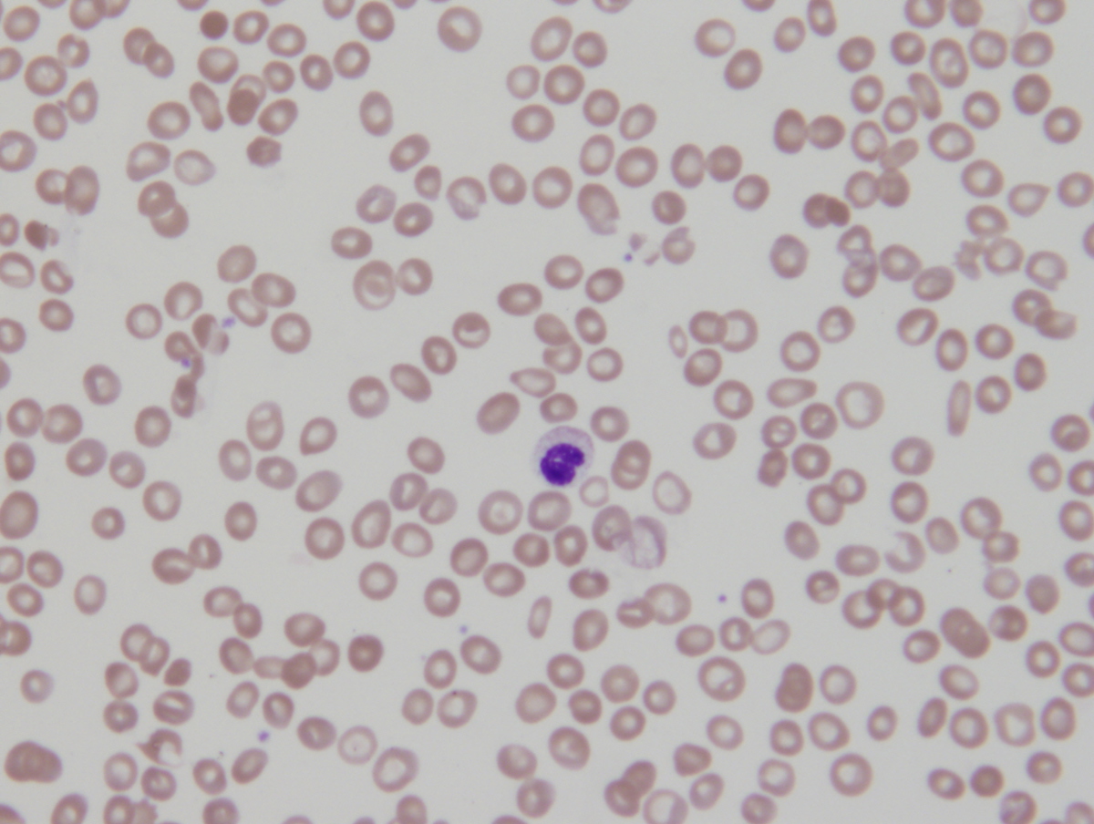 |
| Peripheral blood: dysplastic neutrophils 2 | 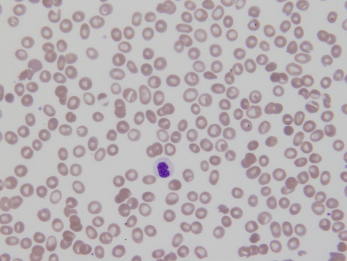 |
| Peripheral blood: circulating blast | 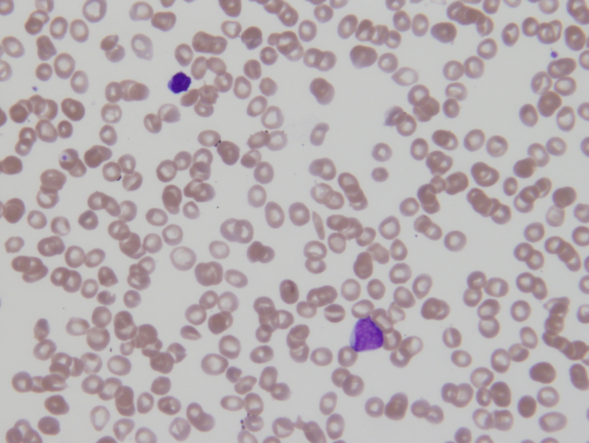 |
| Bone marrow aspirate smear showing blasts | 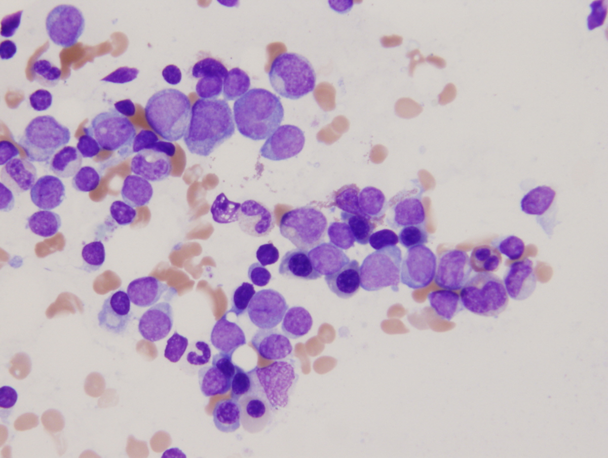 |
| Bone marrow aspirate smear showing blasts 2 | 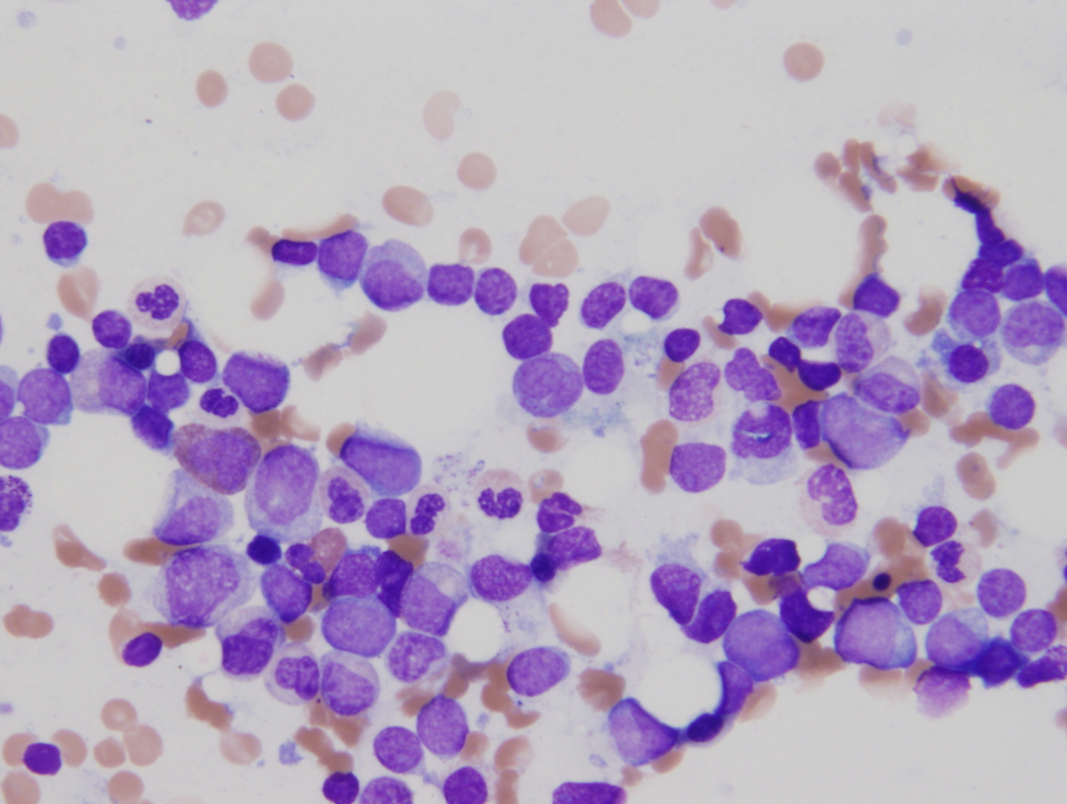 |
| Bone marrow clot section showing increased blasts 1 | 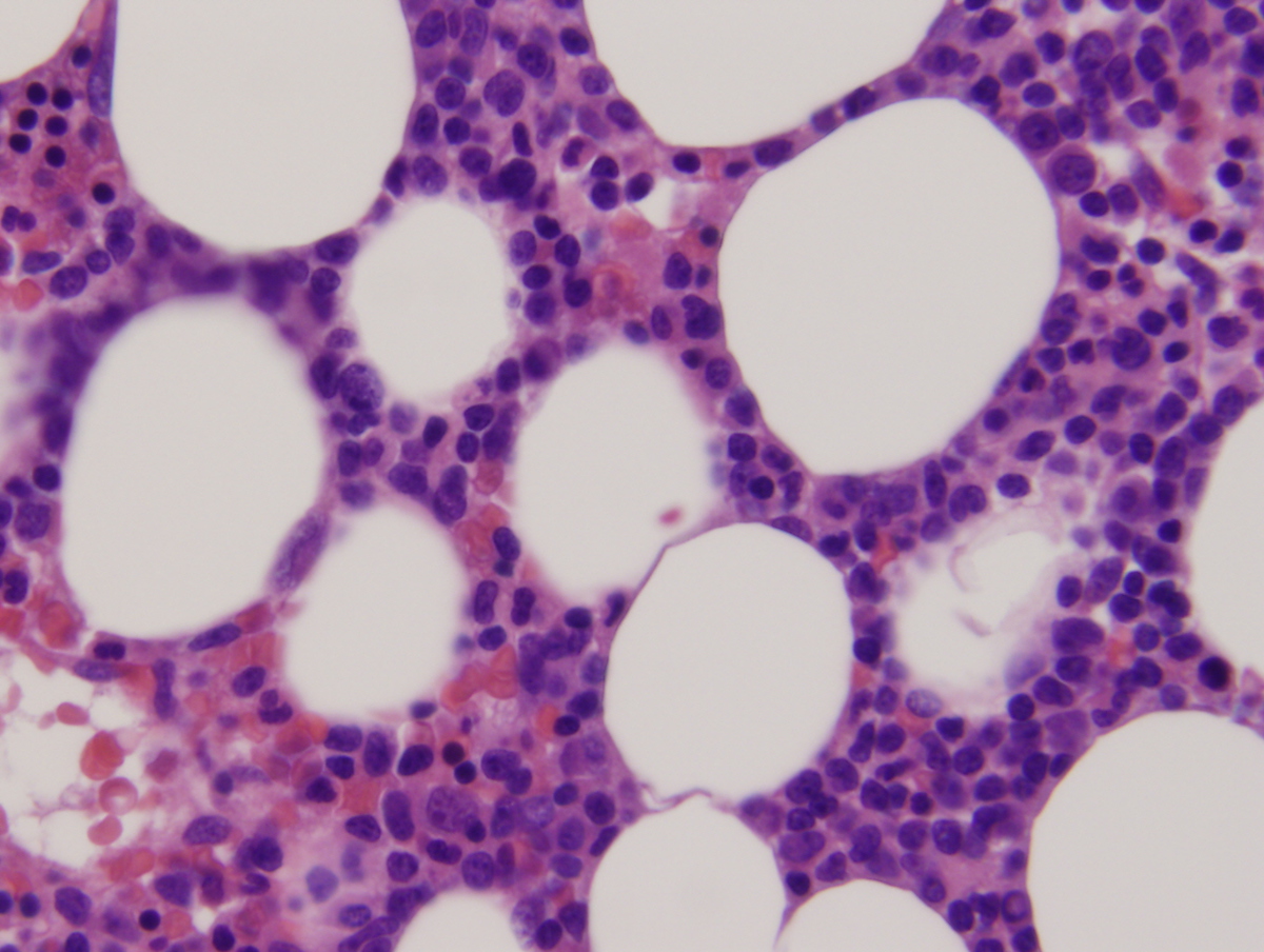 |
| Bone marrow clot section showing increased blasts 2 | 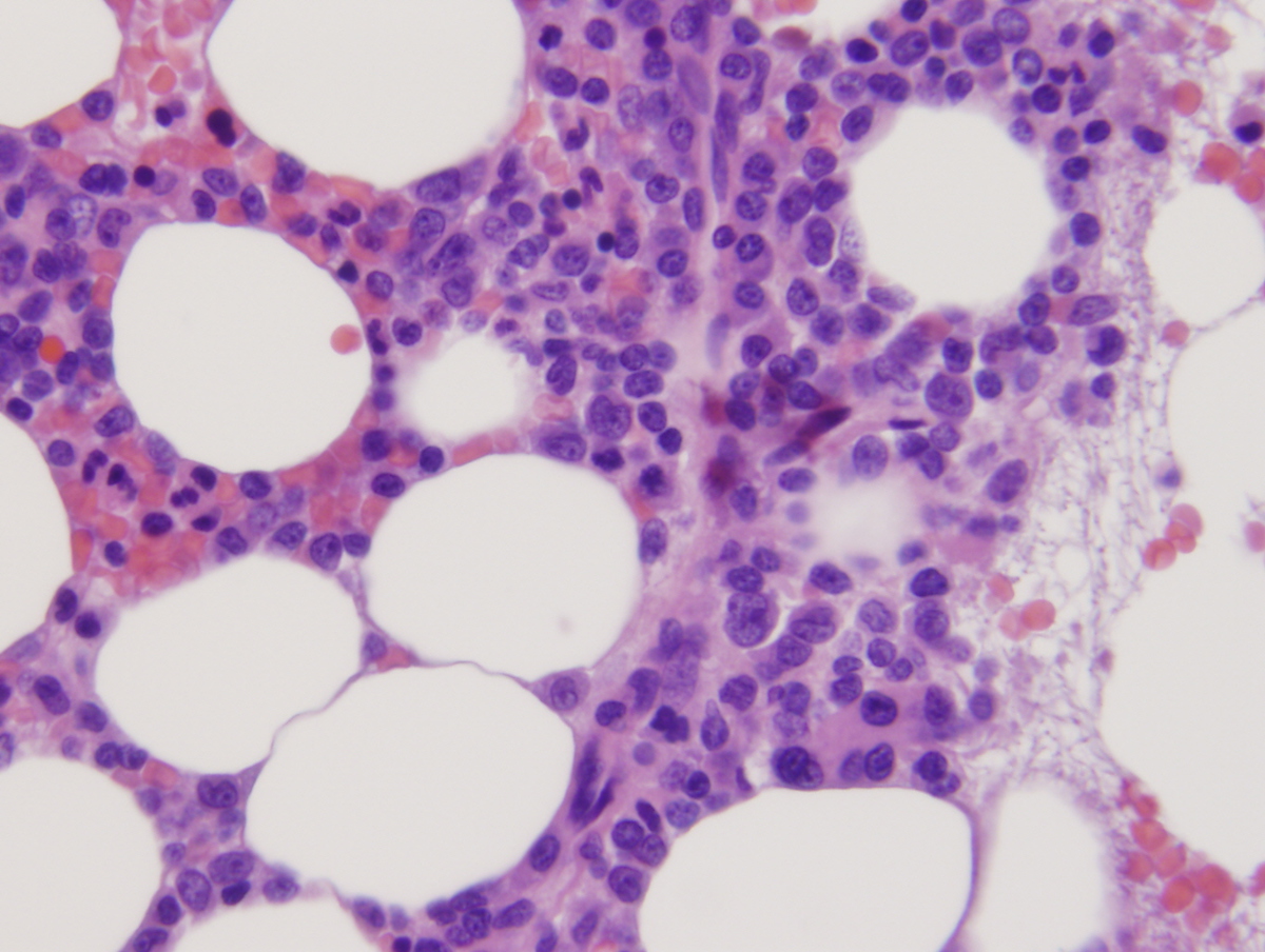 |
| CD34 immunostain highlights blasts | 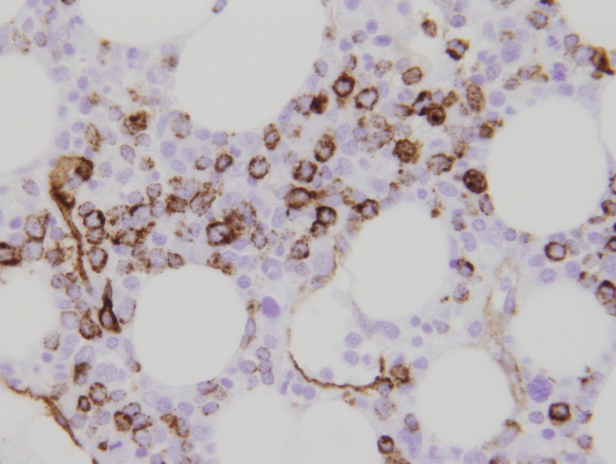 |
| Additional figure 1: H&E (dysplastic megakaryocytes) | 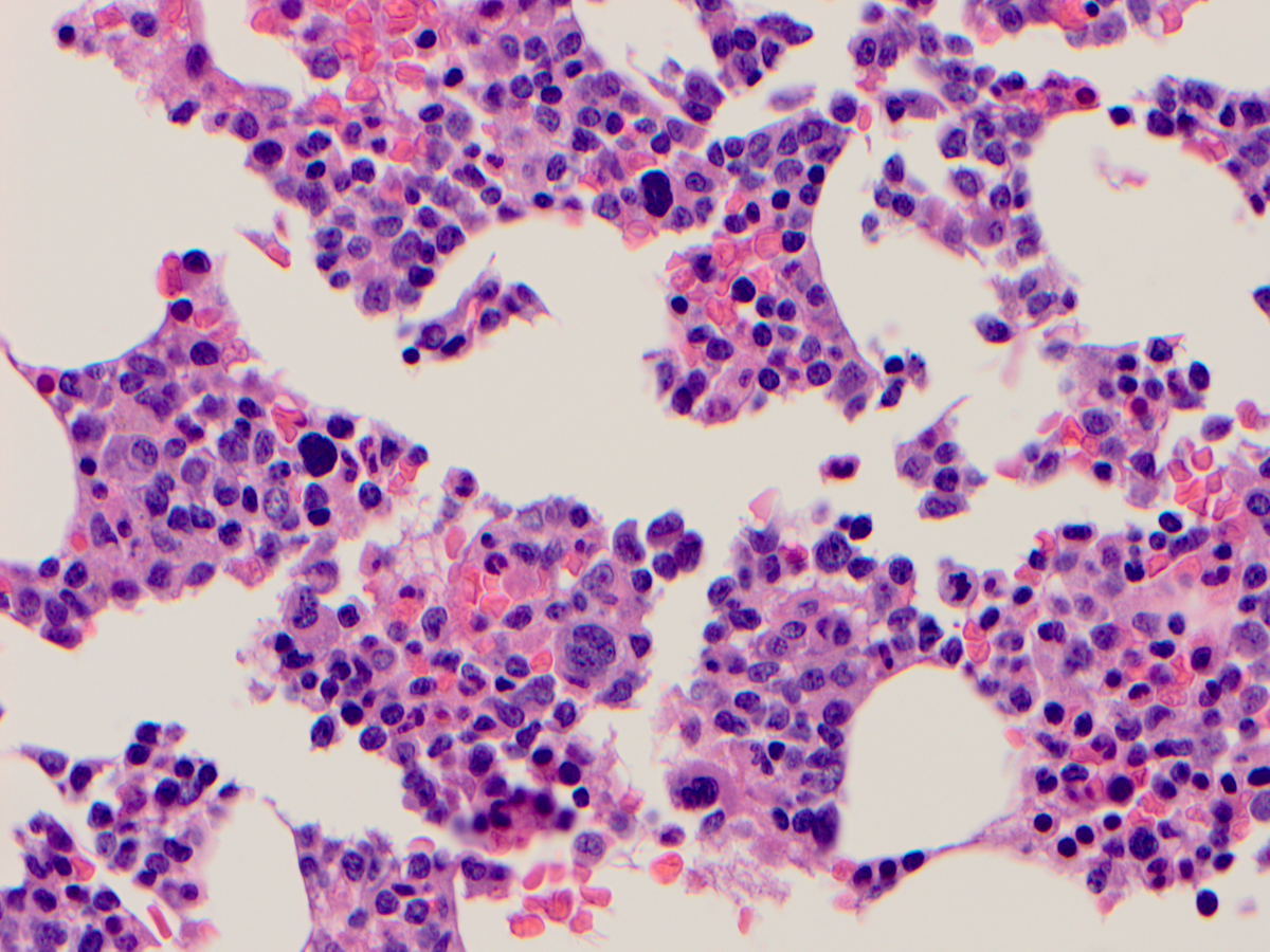 |
| Additional figure 2: CD11c IHC | 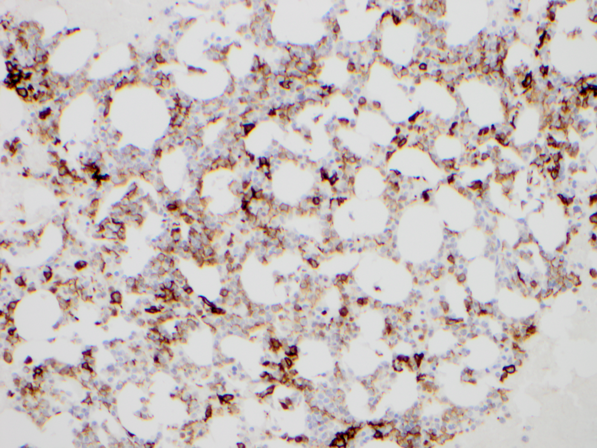 |
| Additional figure 3: Lysozyme IHC | 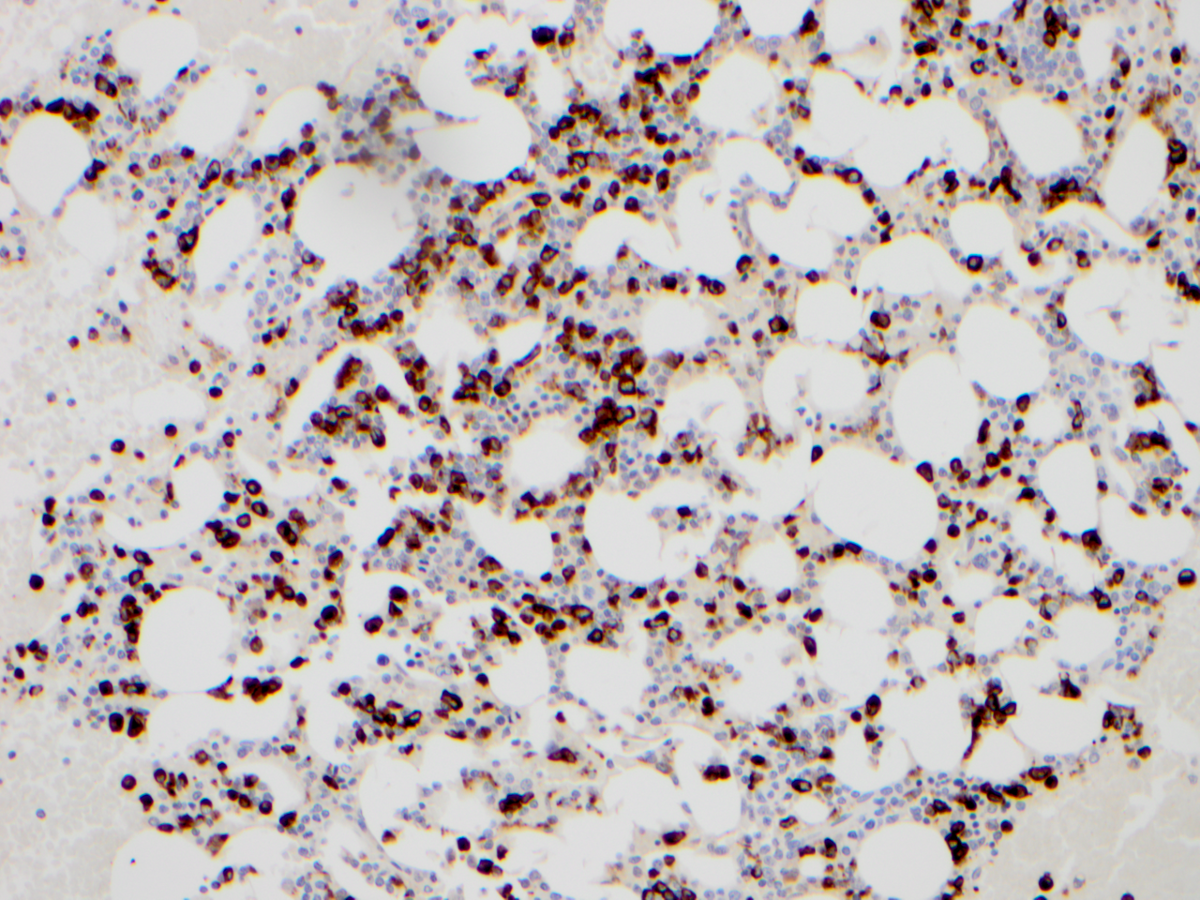 |
| Additional figure 4: MPO IHC | 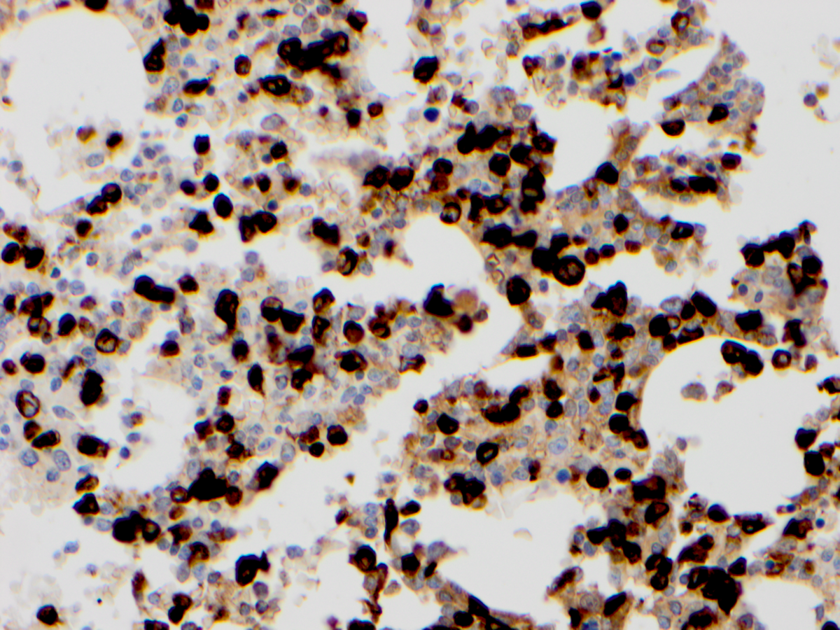 |