Institution: Mayo Clinic Rochester
Additional authors:Dr. Tal Oren (Stamford Pathology Group), Dr. Rhett Ketterling (Mayo Clinic Rochester), and Dr. William Macon (Mayo Clinic Rochester)
Session: T Lymphoblastic Leukemia/Lymphoma
HISTORY
The patient is a 53-year-old male who presented with an enlarged cervical lymph node. No significant history.
DETAILS
The enlarged lymph node is excised with a portion submitted fresh for flow cytometry and cytogenetics. On microscopic examination, the lymph node architecture is near completely effaced by an interfollicular infiltrate of blastic-appearing cells admixed with scattered small lymphocytes. Aggregates of small lymphocytes are noted which represent residual portions of normal lymph node follicles. The blastic-appearing cells are small to intermediate with fine chromatin, inconspicuous nucleoli and scant sytoplasm. Increased mitotic figures are seen.
IMMUNOHISTOCHEMISTRY AND FLOW CYTOMETRY
Immunohistochemical studies were performed on the lymph node. The interfollicular infiltrate is positive for CD3, CD7, CD33, CD34 and CD117. Scattered TdT positive cells are noted. Scattered myeloperoxidase positive cells are noted but are not unequivocally blasts. Flow cytometric immunophenotyping reveals a population of blasts with the following immunophenotypic characteristics: dim CD45+, CD34+, CD117+, cytoplasmic CD3+, CD5 partial +, CD7+, CD33+, CD38+ and HLA-DR partial +. The blasts are negative for CD1a, CD2, surface CD3, CD4, CD8, CD10, CD13, CD14, CD19, CD20, CD56, CD64, cytoplasmic CD79a, cytoplasmic myeloperoxidase and TdT.
CYTOGENETIC FINDINGS
Conventional cytogenetics shows an abnormal karyotype: 46,XY,t(7;12)(p15;p13),del(11)(q13)[2]/46,XY,-13,+14[2]/46,XY[16] FISH studies, performed on the paraffin-embedded tissue, for TLX3/BCL11B, MLLT10/PICALM, TCRB, BCR/ABL, MLL, CDKN2A/D9Z1, TCRAD and STIL/TAL1 are all within normal limits.
MOLECULAR FINDINGS
A T-cell receptor gene rearrangement study is negative.
INTERESTING FEATURES
1) Early T-cell precursor T-lymphoblastic leukemia/lymphoma is a recently recognized (2009) entity with a poor prognosis when treated on contemporary ALL protocols.
2) This entity has a fairly characteristic immunophenotypic profile that is generally distinct from the typical T-lymphoblastic lymphoma/leukemia. (CD34+, CD117+, CD1a(-), CD5 partial +, CD13 or CD33+)
3) Subsequent gene expression profiling studies have shown that the transcriptional profile is similar to that of normal and myeloid leukemia hematopoietic stem cells. This finding raises the possibility that patients may benefit from the addition of myeloid-directed therapies.
4) Recognition of this particular subtype of acute leukemia may be of benefit for patients in terms of therapeutic management.
PROPOSED DIAGNOSIS
T-lymphoblastic leukemia/lymphoma, early T-cell precursor type.
CONSENSUS DIAGNOSIS
T-acute lymphoblastic leukemia/lymphoma, early T-cell precursor immunophenotype
| 10X H&E of lymph node |  |
| 20X H&E of lymph node |  |
| 50X H&E of lymph node |  |
| CD34 |  |
| CD117 |  |
| CD33 |  |
| CD5 | 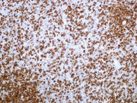 |
| Myeloperoxidase | 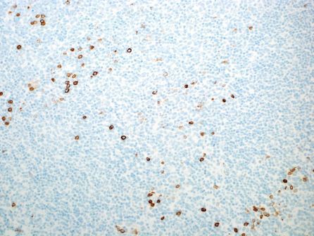 |
| TdT |  |
| Flow cytometry CD34 vs CD117 (population of interest in red) | 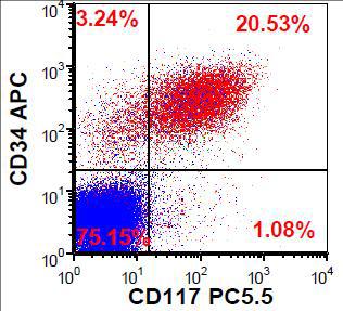 |
| Flow cytometry CD33 vs HLA-DR | 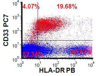 |
| Flow cytometry cytoplasmic CD3 vs CD34 |  |
| Flow cytometry cytoplasmic MPO vs CD34 | 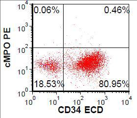 |
| Flow cytometry TdT vs CD34 |  |
| Flow cytometry CD5 vs CD7 |  |
| Flow cytometry CD1a vs CD45 |  |
| Flow cytometry CD13 vs CD14 | 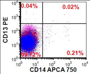 |